Microscope modification could optimize drug testing
Researchers at Purdue University (West Lafayette, IN) have developed a microscope based on concepts of phase-contrast microscopy, which involves using optical devices to view molecules, membranes, or other nanoscale items that may be too translucent to scatter the light involved with conventional microscopes. The development could give doctors a better idea of how safely and effectively a medication will perform in the body.
"One of the problems with using the available microscopes or optical devices is that they require a point of reference for the scattered light, since the object being viewed is too optically transparent to scatter the light itself," explains Garth Simpson, a professor of analytical and physical chemistry in Purdue's College of Science, who led the research team. "We created a unique kind of microscope that stacks the reference object and the one being examined on top of each other with our device, instead of the conventional approach of having them side by side."
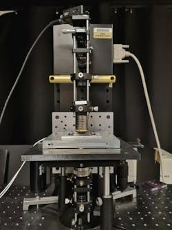
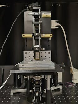
A new type of microscope from Purdue University stacks the reference object and the one being examined on top of each other, instead of the conventional approach of having them side by side. (Image credit: Garth Simpson, Purdue University)
The new microscope uses technology to interfere light from a sample plane and a featureless reference plane, quantitatively recovering the subtle phase shifts induced by the sample. The researchers created their device by adding two small optics to the base design of a conventional microscope. The change allows researchers to gather better information and data about the object being viewed.
"The microscope we have created would allow for better testing of drugs," Simpson says. "You could use our optical device to study how quickly and safely some of the active ingredients in a particular medication dissolve. They may crystallize so slowly that they pass through the body before dissolving, which significantly lowers their effectiveness."
Simpson says that the microscope could also be used for other types of bioimaging, including the ability to study individual cells and membranes from the body for various medical testing.
Full details of the work appear in the journal Optics Express.

