Light microscopy technique images molecules in 3D quickly, with minimal damage to cells
A light microscopy platform developed by Eric Betzig—one of three scientists sharing the 2014 Nobel Prize in Chemistry—and colleagues at the Howard Hughes Medical Institute's Janelia Research Campus can collect high-resolution images rapidly with minimal damage to cells, meaning it can image the 3D activity of molecules, cells, and embryos in fine detail over longer periods than was previously possible.
Related: New light-sheet microscope produces images in real time
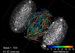
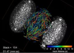
The developers of the new lattice light-sheet microscope have teamed with cell and molecular biologists to produce stunning videos of biological processes across a range of sizes and time scales, from the movements of individual proteins to the development of entire animal embryos.
The new microscope evolved from one that Betzig unveiled in 2011. The Bessel beam plane illumination microscope illuminates samples with a virtual sheet of light, created when a beam of non-diffracting light called a Bessel beam sweeps across the imaging field. It produces high-resolution images with less light damage than a traditional microscope, and is fast enough to record dynamic processes in living cells.
The new microscope operates in two modes. One uses the principles of structured illumination to create very high-resolution images. In this case, the final image is created by collecting and processing multiple images of every plane of the sample. Imaging can be sped up to capture faster processes, albeit at lower resolution, with an alternative "dithered" mode. Light exposure, and thus damage to cells, is lower in the dithered mode; in many cases, tagged proteins are naturally replaced by cells before their signal fades appreciably. "So there are many cells you could look at forever in 3D," Betzig says.
The microscope's high resolution means it can track the movements of individual proteins in 3D, and Betzig says these applications are among the most exciting. "Normally when people do single-molecule studies, they have to do them in thin, flat cells, because the out-of-focus light kills you if it's thick." The lattice's ultra-thin light sheet eliminates that problem and permits single molecules to be seen, even in large multicellular specimens. The microscope is also fast enough to track the rapid growth and retraction of cytoskeletal components in dividing cells, and gentle enough to monitor the molecular dynamics of developmental processes that unfold over many hours.
Betzig wants the lattice light sheet to be widely used, even as technology development continues in his own lab. His team has built a second microscope for Janelia's Advanced Imaging Center, where it will be available to visiting scientists free of charge, and deployed two more of the microscopes to labs at Harvard and the University of California, San Francisco.
Harvard cell biologist Tomas Kirchhausen first used Betzig's lattice light-sheet microscope during a visit to Janelia last year, and knew immediately he wanted to add one to his own lab. After working with Betzig's team to purchase the parts, assemble a "clone" of the original microscope, and set it up at Harvard, Kirchhausen says his team is now generating amazing movies of a variety of dynamic processes in living cells. They are using the technology to track the assembly of thousands of vesicles, watch viruses enter cells, and study how cells change size as they divide. The new microscope has enabled his team to track these processes in a more complete context, since they can now visualize individual molecules within a three-dimensional space, instead of seeing just one plane of a cell at a time, Kirchhausen says.
In fact, Betzig's team freely shares its designs, providing detailed instructions to scientists with the expertise to build their own version of the instrument. Zeiss has licensed the Bessel beam and lattice light-sheet microscopy. "It takes a huge amount of effort to move from a successful high-tech prototype to broader adoption of an imaging technology," Betzig says. "Ultimately, commercialization is the crucial last step to ensuring that these technologies can have broad impact in the research community."
Full details of the work appear in the journal Science; for more information, please visit http://dx.doi.org/10.1126/science.1257998.
-----
Follow us on Twitter, 'like' us on Facebook, connect with us on Google+, and join our group on LinkedIn
Subscribe now to BioOptics World magazine; it's free!

