Optical microscopy helps to identify alcohol abuse damage in neurons at a molecular scale
Researchers at the University of the Basque Country (UPV/EHU; Spain) and the University of Nottingham in England have identified, for the first time, the structural damage caused at a molecular level to the brain by chronic, excessive alcohol abuse. Using a combination of techniques—including optical microscopy—the research team has determined the alterations produced in the neurons of the prefrontal cortex (which controls executive functions such as planning, designing strategies, working memory, selective attention, or control of behavior). The research could lead to new drug developments and therapies that enhance the life of people suffering from alcoholism, as well as reduce morbimortality due to alcoholism.
Related: Laser brain stimulation reduces alcohol consumption in rats
Dr. Luis F. Callado, Dr. Benito Morentin, and Dr. Amaia Erdozain of UPV/EHU, together with Dr. Wayne G. Carter’s team from the University of Nottingham, analyzed the postmortem brains of 20 people diagnosed with alcohol abuse/dependence alongside another 20 non-alcoholic brains. Studying the prefrontal cortex, researchers detected alterations in the neuronal cytoskeleton in the brains of alcoholic patients; in concrete, in the α- and β-tubulin and the β II spectrin proteins. These changes in the neuronal structure, induced by ethanol ingestion, can affect the organization, the capacity for making connections, and the functioning of the neuronal network, and could largely explain alterations in cognitive behavior and in learning attributed to people suffering from alcoholism.
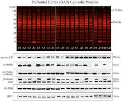
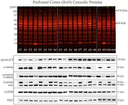
Along with optical microscopy, the researchers used proteomics, Western blot, and mass spectrometry. Optical microscopy demonstrated that the neurons in the prefrontal cortex of the brains of the alcoholic patients had undergone alterations compared to those of non-alcoholic patients. In the following step, the research team used proteomics to identify which proteins were modified in these neurons, which enabled them to determine that the altered elements belong to the families of proteins known as tubulins and spectrins. Tubulins make up the cytoskeleton of neurons, while spectrins are responsible for maintaining cell shape. In this way, both facilitate the relation between and the activity of the components of the brain’s neuronal network.
With the objective of quantifying proteins in each sample, they used the Western blot technique, checking that the levels of proteins were reduced as a consequence of the damage produced by the ethanol. The next stage was using mass spectrometry, which enabled confirming the exact identification of the proteins affected; within the tubulin family, they observed the reduction in the α and β proteins, while amongst the spectrins, they located a decrease in the β II protein.
The research has been published in the journal PLOS One; for more information, please visit http://dx.doi.org/10.1371/journal.pone.0093586.
-----
Don't miss Strategies in Biophotonics, a conference and exhibition dedicated to development and commercialization of bio-optics and biophotonics technologies!
Follow us on Twitter, 'like' us on Facebook, and join our group on LinkedIn
Subscribe now to BioOptics World magazine; it's free!

