Microscopy method has positive implications for muscular dystrophy research
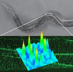
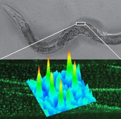
Scientists at the University of Southern California (USC; Los Angeles, CA) have developed a microscopy method that allows them to view single molecules in living animals at high resolution. Importantly, the method enabled them to study a key structural protein of muscle cells to help them understand muscular dystrophy, a degenerative disease.
Related: Optical microscopy enables single-molecule motion capture
Dubbed complementation activated light microscopy (CALM), the method yields imaging resolutions that are an order of magnitude finer than conventional optical microscopy, providing new insights into the behavior of biomolecules at the nanometer scale. The researchers employed CALM to study dystrophin in Caenorhabditis elegans (C. elegans) worms used to model Duchenne muscular dystrophy, the most severe and common form of the disease.
The researchers showed that dystrophin was responsible for regulating tiny molecular fluctuations in calcium channels while muscles are in use. The discovery suggests that a lack of functional dystrophin alters the dynamics of ion channels, helping to cause the defective mechanical responses and the calcium imbalance that impair normal muscle activity in patients with muscular dystrophy.
CALM works by splitting a green fluorescent protein (GFP) from a jellyfish into two fragments that fit together like puzzle pieces. One fragment is engineered to be expressed in an animal test subject, while the other fragment is injected into the animal's circulatory system. When they meet, the fragments unite and start emitting fluorescent light that can be detected with incredible accuracy, offering imaging precisions of around 20 nm.
"Now, for the first time, we can explore the basic principles of homeostatic controls and the molecular basis of diseases at the nanometer scale directly in intact animal models," says Fabien Pinaud, assistant professor at the USC Dornsife College of Letters, Arts and Sciences, who led the work. Scientists from the University Claude Bernard Lyon in France and the University of Würzburg in Germany were also involved in the study.
Next, Pinaud and colleagues will focus on engineering other colors of split-fluorescent proteins to image the dynamics of individual ion channels at neuromuscular synapses within live worms. "It so happens that the same calcium channels we studied in muscles also associate with nanometer-sized membrane domains at synapses where they modulate neuronal transmissions in both normal and disease conditions," Pinaud says. Using multicolor CALM, his team and collaborators will probe how these tiny active zones of neurons are assembled and how they influence the function of calcium channels during neuron activation.
Full details have been published in the journal Nature Communications; for more information, please visit http://dx.doi.org/10.1038/ncomms5974.
-----
Follow us on Twitter, 'like' us on Facebook, and join our group on LinkedIn
Subscribe now to BioOptics World magazine; it's free!

