High-res microscopy helps discover gene essential for sensing joint position
Scientists at The Scripps Research Institute (TSRI; La Jolla, CA) have discovered an important mechanism underlying sensory feedback that guides balance and limb movements. The finding, which they uncovered in Drosophila fruit fly leg joints using high-resolution microscopy, centers on a gene and a type of nerve cell required for detection of leg-joint angles.
If the research team's findings can be fully replicated in humans, they could lead to a better understanding of, as well as treatments for, disorders arising from faulty proprioception (the detection of body position). The proprioceptive sense of how the limbs are positioned is what enables a person, even with eyes closed, to touch the tip of the nose with the tip of a finger—an ability easily impaired by alcohol, which is why traffic police often test suspected drunk drivers this way.
Related: NEUROLOGY/BRAIN IMAGING: A new tool for brain discovery
Scientists have known that proprioceptive signals originate from so-called mechanosensory neurons, whose nerve ends are embedded in muscles, skin, and other tissues. The stretching or compression of these tissues opens ion channels in the nerve membrane, which results in a signal to the brain. What hasn’t been clear is how such a neuron can specialize in sensing just one type of membrane-distorting stimulus—such as the angle of a limb joint—yet exclude others, such as impact pressures.
Boaz Cook, an assistant professor at TSRI who led the work, and colleagues began with a special collection of Drosophila containing a variety of uncataloged mutations. The scientists sifted through the collection looking for mutant flies with walking impairments and soon zeroed in on several impaired walkers that turned out to have mutations in the same gene.
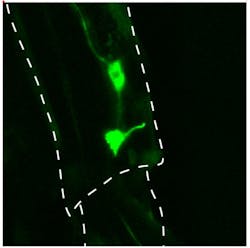
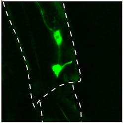
The scientists named the gene stumble (stum for short) for the abnormality caused by its absence. Using a fluorescent tracer, they then localized the expression of stum in normal flies to neurons that lay close to the three main leg joints. Each neuron’s input-sensing tendril (dendrite) grew right up to the joints—a sign that its evolved function is to detect joint angle.
The researchers also found that the protein specified by the stum gene normally migrates to the tip of each dendrite. With high-resolution microscopy, they imaged each of these tips and observed an extra length branching more or less sideways at the joint. At ordinary, at-rest joint angles, the relative positions of the main dendrite tip and its side branch stayed more or less the same; however, at extreme joint angles, the pair stretched out. As they did, the level of calcium ions in the neuron rose sharply, suggesting that ion channels had opened and the neuron was becoming active.
Cook noted the results show how a seemingly general mechanosensory, membrane-stretch-sensitive neuron can evolve a specificity for a particular type of proprioceptive signal. “It’s a nice example of how you can create that specificity from something that only stretches mechanically,” he says.
The team is now trying to nail down the specific role of stum proteins in Drosophila and to determine whether the human version of stum—which has never been characterized—also works in joint angle sensing. Some sensory role for the human version of stum is likely, as the stum gene has been remarkably well conserved throughout animal evolution. Cook and his colleagues were even able to restore some normal walking ability to stum-mutant flies by adding the mouse version of the stum gene. “Stum is probably doing the same thing in all animals,” he said.
Full findings appear in the journal Science; for more information, please visit http://dx.doi.org/10.1126/science.1247761.
-----
Follow us on Twitter, 'like' us on Facebook, and join our group on LinkedIn
Subscribe now to BioOptics World magazine; it's free!

