Tricked-out iPhone performs microscopy and spectroscopy
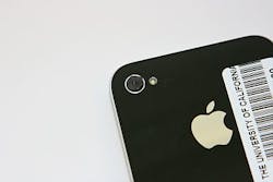
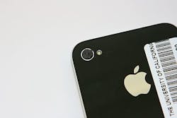
Seeking a way to provide a cheaper medical imaging device using materials that cost as much as a typical smartphone app, researchers from the University of California, Davis (UC Davis; Davis, CA) have transformed an iPhone into a device able to perform detailed microscopy and spectroscopy. The iPhones could help doctors and nurses diagnose blood diseases in developing nations where many hospitals and rural clinics have limited or no access to laboratory equipment, enabling them to transmit real-time data to colleagues around the globe for further analysis and diagnosis.
For example, field workers could put a blood sample on a slide, take a picture and send it to specialists to analyze, says Sebastian Wachsmann-Hogiu, a physicist with UC Davisâ Department of Pathology and Laboratory Medicine and the Center for Biophotonics, Science and Technology, and lead author of the research.
To build the microscope's lens, the team used a 1-mm-diameter ball lens that costs $30â40 USD in their prototype, but mass-produced lenses could be substituted to reduce the price. Kaiqin Chu, a post-doctoral researcher in optics, inserted the ball lens into a hole in a rubber sheet, and then simply taped the sheet over the smartphoneâs camera. At 5x magnification the ball lens is as powerful as a simple magnifying glass, but when paired with a smartphone camera it could resolve features as small as 1.5 µmâsmall enough to identify different types of blood cells.
How does low magnification produce such high-resolution images? First, ball lenses excel at gathering light, which determines resolution. Second, the cameraâs semiconductor sensor consists of millions of light-capturing cells, with each cell measuring about 1.7 µm across. This is small enough to capture precisely the tiny high-resolution image that comes through the ball lens.
But the curvature a ball lens' sphere bends light as it enters the ball, distorting the image, except for a very small spot in the center. The researchers used digital image processing software to correct for this distortion. They also used the software to stitch together overlapping photos of the tiny in-focus areas into a single image large enough for analysis.
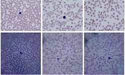
Even though smartphone micrographs are not as sharp as those from laboratory microscopes, they are able to reveal important medical information, such as the reduced number and increased variation of cells in iron deficiency anemia, and the banana-shaped red blood cells characteristic of sickle cell anemia.
Wachsmann-Hogiuâs team is working with UC Davis Medical Center to validate the device and determine how to use it in the field. They may also add features, such as larger lenses to diagnose skin diseases and software to count and classify blood cells automatically in order to provide instant feedback and perhaps recognize a wider range of diseases.
When researchers need additional diagnostic tools, the microscope could be swapped for a simple spectrometer that also uses light collected by the iPhoneâs camera. Like the microscope, the iPhoneâs spectrometer takes advantage of smartphone imaging capabilities. The spectrometer that the researchers added to the iPhone starts with a short plastic tube covered at both ends with black electrical tape. Narrow slits cut into the tape allow only roughly parallel beams of light from the sample to enter and exit the tube to spread the light into a spectrum of colors to then identify various molecules. Though the spectrometer is still in its early stages, the researchers believe it could measure the amount of oxygen in the blood and help diagnose chemical markers of disease.
The research team will present their findings at the Optical Societyâs (OSA) Annual Meeting, Frontiers in Optics (FiO) 2011, taking place in San Jose, CA, from October 16â20. The presentation, âMicroscopy and Spectroscopy on a Cell Phone,â will take place Wednesday, October 19, at 12 p.m. at the Fairmont San Jose Hotel.
-----
Follow us on Twitter, 'like' us on Facebook, and join our group on LinkedIn
Follow OptoIQ on your iPhone; download the free app here.
Subscribe now to BioOptics World magazine; it's free!


