Digital holographic microscopy observes neuron activity in 3-D
A team of researchers from École Polytechnique Fédérale de Lausanne (EPLF) and Centre Hospitalier Universitaire Vaudois (CHUV; both of Lausanne, Switzerland) used digital holographic microscopy (DHM) to observe neuronal activity in real-time and in 3-D, with up to 50 times greater resolution than ever before. Published recently in the Journal of Neuroscience, DHM shows promise in testing out new drugs to fight neurodegenerative diseases such as Alzheimer's and Parkinson's.
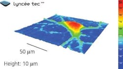
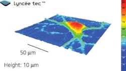
Pierre Magistretti of EPFL's Brain Mind Institute, also a lead author of the paper, explains that because DHM is noninvasive, it allows for extended observation of neural processes without the need for electrodes or dyes that damage cells. The method gives important information about the shape, dynamics and activity of neurons and creates 3-D navigable images down to 10 nm precision, adds senior team member Pierre Marquet.
DHM works by pointing a single-wavelength laser beam at the neurons, collecting the distorted wave on the other side and then comparing it to a reference beam. A computer then numerically reconstructs a 3-D image of the neurons using an algorithm developed by the authors. In addition, the laser beam travels through the transparent cells and important information about their internal composition is obtained.
Normally applied to detect minute defects in materials, Magistretti, along with DHM pioneer and EPFL professor in the Advanced Photonics Laboratory, Christian Depeursinge, decided to use DHM for neurobiological applications. In the study, their group induced an electric charge in a culture of neurons using glutamate, the main neurotransmitter in the brain. This charge transfer carries water inside the neurons and changes their optical properties in a way that can be detected only by DHM. Thus, the technique accurately visualizes the electrical activities of hundreds of neurons simultaneously, in real-time, without damaging them with electrodes, which can only record activity from a few neurons at a time.
Without the need to introduce dyes or electrodes, DHM can be applied to high content screeningâthe screening of thousands of new pharmacological molecules. This advance has important ramifications for the discovery of new drugs that combat or prevent neurodegenerative diseases such as Parkinson's and Alzheimer's, since new molecules can be tested more quickly and in greater numbers. Magistretti notes that what normally would take 12 hours in the lab can now be done in 15 to 30 minutes, greatly decreasing the time it takes for researchers to know if a drug is effective or not.
The success of DHM for high content screening has already resulted in a start-up company at EPFL called LyncéeTec.
-----
Follow us on Twitter, 'like' us on Facebook, and join our group on LinkedIn
Follow OptoIQ on your iPhone; download the free app here.
Subscribe now to BioOptics World magazine; it's free!

