Confocal microscopy method effective as diagnostic tool for sarcoidosis
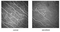
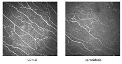
An international team of researchers from Leiden University Medical Center (Leiden, The Netherlands) and the University of Manchester (Manchester, England) used a corneal confocal microscope to photograph nerve fibers in layers of the cornea followed by computerized image analysis to quantify and predict severity of the symptoms of painful small fiber neuropathy in patients with sarcoidosis (growth of tiny collections of inflammatory cells in the eyes, as well as other parts of the body).
Related: Super-res microscopy yields clearest images of immune cells
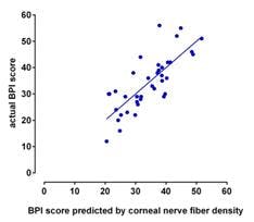
Using their method, the researchers discovered that corneal nerve fiber number and length correlate strongly with intra-epidermal nerve fiber density obtained by skin biopsy from the leg, as well as correlate strongly with the score of a patient questionnaire on symptoms of neuropathy. The cornea has the highest density of nerve fibers of any tissue in the body, and repeat measurements without discomfort to the patient can be used to gain significantly more information on nerve fiber damage. The correlation identified using the microscopy method with patient-reported symptoms provides a rapid and simple way of diagnosing neuropathy resulting from small nerve fiber loss and damage in sarcoidosis patients, and also offers a way to objectively evaluate the effects of therapeutic interventions on a notoriously difficult disease to follow.
âWe were quite surprised and pleased to discover that by using corneal confocal microscopy, the amount of useful information on the patientâs neuropathic condition could be obtained that surpasses other diagnostic methods, even those that are much more traumatic for the patient. Additionally, we can essentially diagnose disease objectively, without relying on subjective information provided by the patient,â explains Dr. Anthony Cerami, senior author and CEO of Araim Pharmaceuticals (Ossining, NY), which funded the work. âCurative therapies for small nerve fiber loss and damage are lacking, and we anticipate that use of this technique will permit us and others to assess therapeutics that promote nerve regrowth in the cornea that is reflective of nerve regrowth in other target tissues of neuropathy.â
Dr. Michael Brines, lead author, described the clinical study conducted at Leiden University Medical Center in which the correlation was identified. Sarcoidosis patients were screened for corneal nerve fiber density and then re-evaluated after 28 days, without any change to their medication regimen, as part of a larger study that evaluated a therapeutic intervention, the results of which will be reported at a later date. Dr. Brines commented, âWe anticipate that once the correlation between corneal nerve fiber status and objective measurements is firmly established, this non-invasive method can be used as a valid surrogate endpoint for small fiber neuropathy. The holy grail of therapeutic intervention in chronic disease is disease modification, and if the correlation holds up, the eye provides a lens, so to speak, on whatâs going on in tissues affected by the disease process and responsible for the poor quality of life of its victims.â
Full details of the research team's work appear in the journal Technology; for more information, please visit http://www.worldscientific.com/doi/abs/10.1142/S2339547813500039.
-----
Follow us on Twitter, 'like' us on Facebook, and join our group on LinkedIn
Subscribe now to BioOptics World magazine; it's free!


