Modified mobile phone camera becomes mini microscope for cheap diagnostics
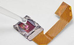
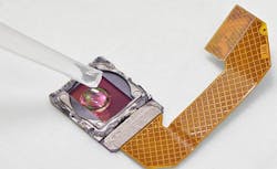
Researchers at the Institute for Molecular Medicine Finland (FIMM) at the University of Helsinki and Karolinska Institutet (Solna, Sweden), by modifying a mobile phone's camera, have shown that novel techniques for high-resolution biomedical imaging and image transfer over data networks may be utilized to diagnose tropical parasitic infections.
Related: Algorithms and cell phone add-on enable powerful, inexpensive microscopy
Related: Tricked-out iPhone performs microscopy and spectroscopy
Dr. Johan Lundin and Dr. Ewert Linder led the research team, who modified inexpensive imaging devices such as a webcam selling for $13 (â¬10) and a mobile phone camera into a mini microscope. The test sample was placed directly on the exposed surface of the image sensor chip after removal of the optics. The resolution of such mini microscopes was dependent on the pixel size of the sensor, but sufficient for identification of several pathogenic parasites.
In a study, the researchers were able to use the mini microscopes they constructed to yield images of parasitic worm eggs present in urine and stools of infected individuals. They first utilized their approach to detect urinary schistosomiasis, a severely under-diagnosed infection that affects hundreds of millions of people, primarily in sub-Saharan Africa. For diagnostics at the point-of-care, they developed a highly specific pattern recognition algorithm that analyzes the image from the mini-microscope and automatically detects the parasite eggs.
"The results can be exploited for constructing simple imaging devices for low-cost diagnostics of urogenital schistosomiasis and other neglected tropical infectious diseases," says Lundin. "With the proliferation of mobile phones, data transfer networks, and digital microscopy applications, the stage is set for alternatives to conventional microscopy in endemic areas."
Full details of the work appear in the journal PLOS Neglected Tropical Diseases; for more infromation, please visit http://www.plosntds.org/article/info%3Adoi%2F10.1371%2Fjournal.pntd.0002547.
-----
Follow us on Twitter, 'like' us on Facebook, and join our group on LinkedIn
Subscribe now to BioOptics World magazine; it's free!

