Stimulated Raman scattering could help boost accuracy of brain tumor surgery
A team of University of Michigan Medical School (Ann Arbor, MI) and Harvard University (Cambridge, MA) researchers are turning to stimulated Raman scattering (SRS) to enable much more accurate brain tumor surgery. Combined with microscopy, the technique could allow surgeons to distinguish cancer tissue from normal brain tissue at the microscopic level while they are operating, and avoid leaving behind cells that could spawn a new tumor.
Related: Miniature microscopes for guiding brain tumor resection
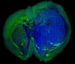
They used the SRS microscopy technique to distinguish tumor from healthy tissue in the brains of living mice—and then showed that the same was possible in tissue removed from a patient with glioblastoma multiforme, one of the most deadly brain tumors.
Now, the team is working to develop the technique for use during an operation to guide them in removing tissue, and test it in a NIH-funded clinical trial at the University of Michigan (U-M).
"Though brain tumor surgery has advanced in many ways, survival for many patients is still poor, in part because surgeons can't be sure that they've removed all tumor tissue before the operation is over," says co-lead author Daniel Orringer, MD, a lecturer in the U-M Department of Neurosurgery who has worked with the Harvard team since a chance meeting with a team member during his U-M residency.
In the SRS technique, they can detect a weak light signal that comes out of a material after it is hit with light from a noninvasive laser. By carefully analyzing the spectrum of colors in the light signal, the researchers can tell a lot about the chemical makeup of the sample.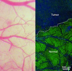
Over the past 15 years, Xiaoliang Sunney Xie, Ph.D., of the Department of Chemistry and Chemical Biology at Harvard University—the senior author of the new paper—has advanced the technique for high-speed chemical imaging. By amplifying the weak Raman signal by more than 10,000 times, it is now possible to make multicolor SRS images of living tissue or other materials. The team can even make 30 new images every second—the rate needed to create videos of the tissue in real time.
"Biopsy has been the gold standard for detecting and removing these types of tumors," says Xie, the Mallinckrodt Professor of Chemistry and Chemical Biology. "But this technique, we believe, is better because it's live. Surgeons can now skip all the steps of taking a biopsy, freezing and staining the tissue--this technique allows them to do it all in vivo."
The work marks the first time that SRS microscopy has been used in a living organism to see the "margin" of a tumor (the boundary area where tumor cells infiltrate among normal cells), which is the hardest area for a surgeon to operate—especially when a tumor has invaded a region with an important function. The technique can distinguish brain tumor from normal tissue with remarkable accuracy by detecting the difference between the signal given off by the dense cellular structure of tumor tissue, and the normal, healthy gray and white matter. Better still, the authors suggest that SRS microscopy may be as accurate for detecting tumor as the current H&E staining approach used in brain tumor diagnosis.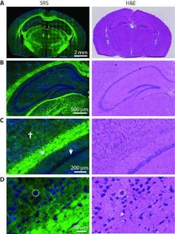
The paper contains data from a test that pitted H&E staining directly against SRS microscopy. Three surgical pathologists, trained in studying brain tissue and spotting tumor cells, had nearly the same level of accuracy, no matter which images they studied. But unlike H&E staining, SRS microscopy can be done in real time, and without dyeing, removing, or processing the tissue.
The current SRS microscopy system is not yet small or stable enough to use in an operating room. The team is collaborating with a start-up company formed by members of Xie's group, called Invenio Imaging (Boston, MA), which is developing a laser to perform SRS through inexpensive fiber-optic components. The team is also working with AdvancedMEMS (San Francisco, CA) to reduce the size of the probe that makes the images possible. And a validation study, to examine tissue removed from consenting U-M brain tumor patients, may begin as soon as next year.
Full details of the work appear in the journal Science Translational Medicine; for more information, please visit http://stm.sciencemag.org/content/5/201/201ra119.