Single VECSEL light source enables multimodal bioimaging
Researchers at Yale University (New Haven, CT) have developed a vertical external-cavity surface-emitting laser (VECSEL) system that acquires two types of microscopy images with a single light source. In a proof-of-concept demonstration of the system's potential for bioimaging, the researchers used it to dynamically image the blood flow and miniscule structural changes of a tadpole embryo's beating heart.
Related: Multimodal imaging system for cancer detection is smaller and cheaper
Although others have achieved multimodal imaging using different light sources, it has been challenging for these systems to maintain optical alignment during switching and to adjust for mismatches in illumination intensity. But the Yale research team's laser system design allows switching back and forth between different operating modes with essentially the same beam, keeping the direction and optical powers similar so that everything stays well aligned, explains Hui Cao of Yale's Applied Physics Laboratory, who developed the laser system.
The new light source grew out the researchers' work to develop a laser that could eliminate the speckle pattern that obscures images acquired using laser light. Speckle is an optical artifact created by lasers' high spatial coherence—the fact that the light waves emitted from a laser are all synchronized, or in step with each other.
Illuminating a sample with a non-coherent light, such as that from LEDs or a white lamp, doesn't cause speckle. However, these light sources aren't bright enough for the fast imaging speeds, and thus short exposure times, required to follow dynamic biomedical processes such as a beating heart.
In developing their new system, Cao and colleagues took advantage of the fact that the particular pattern of speckle created by a sample can provide information about the dynamics of the cells scattering the light. Since the 1990s, a method called laser speckle contrast imaging has used the temporal change of speckle to provide information about blood flow.
The new laser system can create light with low spatial coherence for speckle-free structural images or be quickly switched to high spatial coherence for speckle contrast imaging of the same tissue.

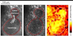
The researchers conducted an initial demonstration of their light source by imaging a beating heart from a Xenopus tadpole, an animal model that Michael Choma, coauthor of the research team's paper describing their work, is studying to better understand heart disease. The researchers first acquired low-coherence images to show the heart's structure and how it changes during a heart beat. They then switched to high coherence to measure the flow of blood through the heart using laser speckle contrast imaging. Choma says that having straightforward ways to assess heart function in animal models such as Xenopus allows them to understand the effects of a gene mutation related to congenital heart disease or to study the impact of a drug on embryo heart function. The light source may someday also be useful for clinical applications that involve visualizing and quantifying blood flow in the retina or during certain surgeries.
The researchers' light source features a very simple design of only five optical elements, plus a VECSEL. The VECSEL emits light from multiple sites, with the light from each site slightly out of step with all the others. The researchers used all 1000 independent modes of light coming from the VECSEL to generate low-coherence light. By adding a pinhole to the laser cavity, they were able to concentrate all the laser power into just a couple of modes to create high-coherence light for laser speckle contrast imaging. What's more, the system's optical setup is compact, easy to assemble, and inexpensive, as it uses commercially available, off-the-shelf components, Cao says.
Next, the researchers plan to refine the system by using a type of digital mirror array called a spatial light modulator to switch between low and high coherence many times during one heartbeat. This would allow almost simultaneous imaging of blood flow and structure.
Full details of the work appear in the journal Optica; for more information, please visit http://dx.doi.org/10.1364/optica.3.000403.