Intravital microscopy helps discover key mechanism in immune response
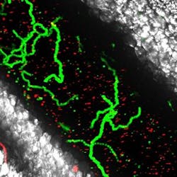
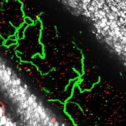
Seeking to understand the role of white blood cells during immune response, researchers in the Pathology and Immunology Department of the Faculty of Medicine at the University of Geneva (UNIGE; Switzerland) used intravital microscopy to follow cell activity in the blood in real time. By doing so, they observed that when resting, the endothelium (blood vessel) produces a protein called CCN1 that coats the internal side of the blood vessel.
Related: Super-resolution microscopy reveals more on how the immune system is regulated
If this protein's activity is blocked, all the patrolling work carried out by the resident monocytes is disrupted, according to Yalin Emre, co-author of the study. "They then move around much slower along the blood vessel wall and fail to check all the necessary areas. We therefore discovered that CCN1 provides a molecular support, allowing resident monocytes to move around efficiently," he explains.
When a virus attacks, an inflammation appears on the affected area and triggers immune response within the body—white blood cells (such as inflammatory monocytes and neutrophils) move quickly to the inflamed area. The previous understanding was that neutrophils were the first defenders to arrive, but the UNIGE research team discovered that neutrophils depend on a group of patrolling monocytes, referred to as "residents," and also on CCN1 proteins, which are produced by blood platelets and by the endothelium. Without the latter, the defenders are not recruited to fight off the virus. So, this discovery could lead to new possible theories regarding antiviral treatments.
When attacked, white blood cells leave the blood circulation and migrate into the tissue around the inflamed area. Neutrophils are the first to be recruited within a few hours, followed by inflammatory monocytes that appear as a backup some time later. Neutrophils are therefore picked up in the area where the endothelium is stressed by the attack. They stick to its wall and then migrate outside the vessel to reach the damaged tissue and to fight against the infection. When resting, the endothelium is incessantly scanned by resident monocytes in charge of patrolling the smallest part of blood vessels to check that all is running smoothly.
In their study, the researchers created blood vessel inflammation using an agent mimicking a viral infection on mice, and observed that the amount of CCN1 bound to the endothelium quadrupled in 20 minutes, the number of patrolling monocytes tripled between 30 and 60 minutes, and neutrophils arrived to the inflamed area 120 minutes after the viral attack started. "By blocking the CCN1 activity during the inflammation, we noticed that the recruitment of patrolling monocytes and neutrophils stopped, which confirms the importance of this protein's role during the early steps of inflammation," Emre explains.
It was necessary still to explain how the CCN1 amount quadrupled so quickly after injury. With the intravital microscopy technique, Emre says, the research team saw that patrolling monocytes usually have no contact with the platelets at rest—therefore, platelets flow freely in the blood. But once inflammation occurs, platelets directly interact with the patrolling monocytes and release the CCN1 protein, which binds onto the endothelium. "The increase of CCN1 amount is essential both for the recruitment of resident monocytes and for their patrolling activity. In the event of a viral attack and if there are no platelets in the blood, the CCN1 level will not rise, therefore abolishing any recruitment of patrolling monocytes that cannot call upon either neutrophils or inflammatory monocytes," he adds.
CCN1 is therefore essential to both guide the patrollers and to trigger the immediate immune response in the event of a viral attack. This knowledge is promising for new possible therapies for antiviral treatment, as previous research focused mainly on the cells (lymphocytes, NK cells, neutrophils, and macrophages) detected in the infected tissues to attempt fighting off the virus.
Full details of the work appear in the Proceedings of the National Academy of Sciences; for more information, please visit http://dx.doi.org/10.1073/pnas.1607710113.

