SERRS nanoprobes in development could enable tumor visualization in vivo
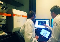
The Kircher laboratory at Memorial Sloan Kettering Cancer Center (MSKCC; New York, NY) is developing novel nanoprobes for molecular imaging, image-guided therapy, and theranostics. The research team's ultimate goal is to develop a universal technology that allows precise determination of the actual spread of a tumorin vivo. Currently, surgeons cannot see the microscopic extent of the tumor during a procedure, which is essential information for tumor removal and avoiding excess tissue excision.
Related: Handheld Raman scanner could assist in complete brain tumor removal
Physician-scientist Dr. Moritz Kircher, a member of the Department of Radiology, the Center for Molecular Imaging and Nanotechnology, and the Brain Tumor Center at MSKCC, is working on a new generation of nanometer-sized imaging beacons. These allow detection, during surgeries and minimally invasive procedures, of the macroscopic extent of the primary tumor and its true microscopic spread, as well as information on satellite micrometastases. These nanobeacons can be located using surface-enhanced resonance Raman scattering (SERRS), a highly sensitive technique that uses a resonance Raman effect and combines with a surface enhancement obtained from nanoparticles to give dramatic enhancements to the normally weak Raman signal. SERRS can work with other whole-body imaging methods like magnetic resonance imaging (MRI) or positron emission tomography (PET).
Kircher explains that his research team's SERRS nanoparticles are not only universal with regards to tumor type, they also allow them to detect microscopic tumor extensions from the main tumor into the periphery, microscopic loco-regional metastases, and even premalignant lesions.
Recent details of the work appear in the Journal of Nuclear Medicine; for more information, please visit http://dx.doi.org/10.2967/jnumed.115.158196.