Computational approach enables 'stainless' staining for optical imaging

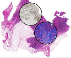
Researchers at the University of Illinois at Urbana-Champaign have developed a new approach to histology based on using infrared (IR) microspectroscopy. Instead of using stains, the spectra measure the chemical constitution of cells and tissues directly.
Related: Computational microscopy approach can 'paint' tissue samples with light
By using computational techniques, the researchers—led by Rohit Bhargava, a professor in the university's Bioimaging Science and Technology Group—were able to relate spectral properties to known staining patterns of tissue. The outcome is that that molecular stains can be reproduced without staining the tissue, but by using the intrinsic molecular contrast of the tissue and computation. That means any sample can be stained for desired stains without material cost, time, or effort while leaving precious tissue pristine for downstream analyses.
Another use of the approach can be in the analysis of small amounts of samples—for example, from a thin needle biopsy. In cases where materials are limited or there may be a need to closely correlate multiple expressed molecules, it may not be possible to obtain multiple samples from the same biopsy for multiple stains. The method developed in this study could be a solution, allowing the user to simply "dial-in" a required stain. The study is timely, as it builds on the emergence of chemical imaging and maturation of computation from the sciences/engineering side and the drive to greater molecular content from the biomedical/clinical side. The development of this approach promises to have immediate and long-term impact in changing pathology to a multiplexed molecular science—in both research and clinical practice.
Full details of the work appear in the journal Technology; for more information, please visit http://dx.doi.org/10.1142/S2339547815200010.
-----
Follow us on Twitter, 'like' us on Facebook, connect with us on Google+, and join our group on LinkedIn