Smartphone video microscope detects parasites in blood within 2 minutes
Researchers at the University of California-Berkeley (UC Berkeley) have developed a new smartphone microscope that uses video to automatically detect and quantify infection by parasitic worms in a drop of blood. This next generation of UC Berkeley's CellScope technology could help revive efforts to eradicate debilitating filarial diseases in Africa by providing critical information to health providers in the field.
Related: Infectious disease control with portable CMOS-based diagnostics
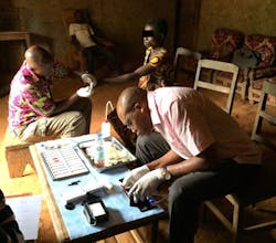
The research team—led by Daniel Fletcher, an associate chair and professor of bioengineering whose lab pioneered the CellScope—teamed up with Dr. Thomas Nutman from the National Institute of Allergy and Infectious Diseases (NIAID), and collaborators from Cameroon and France to develop the device. They conducted a pilot study in Cameroon, where health officials have been battling the parasitic worm diseases onchocerciasis (river blindness) and lymphatic filariasis.
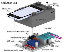
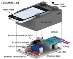
The video CellScope, which uses motion instead of molecular markers or fluorescent stains to detect the movement of Loa loa worms, was as accurate as conventional screening methods, the researchers found. The mobile phone microscope, named CellScope Loa, pairs a smartphone with a 3D-printed plastic base where the sample of blood is positioned. The base included LED lights, microcontrollers, gears, circuitry, and a USB port.
Control of the device is automated through an app the researchers developed for this purpose. With a single touch of the screen by the healthcare worker, the phone communicates wirelessly via Bluetooth to controllers in the base to process and analyze the sample of blood. Gears move the sample in front of the camera, and an algorithm automatically analyzes the telltale "wriggling" motion of the worms in video captured by the phone. The worm count is then displayed on the screen.
Fletcher says previous field tests revealed that automation helped reduce the rate of human error. The procedure takes about two minutes or less, starting from the time the sample is inserted to the display of the results. Pricking a finger and loading the blood onto the capillary adds another minute to the time. The short processing time allows health workers to quickly determine on site whether it is safe to administer the antiparasitic drug ivermectin (IVM).
The researchers are now expanding the study of CellScope Loa to about 40,000 people in Cameroon.
Full details of the work appear in the journal Science Translational Medicine; for more information, please visit http://dx.doi.org/10.1126/scitranslmed.aaa3480.
-----
Follow us on Twitter, 'like' us on Facebook, connect with us on Google+, and join our group on LinkedIn


