Research team uses Raman spectroscopy to study various childhood diseases
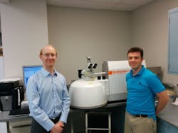
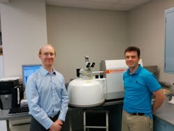
Researchers at the Children's Hospital of Michigan and Wayne State University (both in Detroit, MI) used Raman spectroscopy—specifically, confocal Raman microscopy—to help them study various diseases, with particular focus on childhood diseases.
Related: Spectral imaging may improve surgery for colon disease in infants
One of the diseases the team studied is Hirschsprung's disease, a bowel disease. The objective of Brady King, Ph.D., a research associate at the Children's Hospital of Michigan and an adjunct assistant professor of surgery at Wayne State University, and colleagues is to develop Raman spectroscopy for use in clinical applications. The ultimate goal is to have this type of technology available in the operating room for accurate, real-time diagnosis of tissue during operations.
King explains that Raman spectroscopy is well suited for a pathological workflow, as its accurate staging and imaging have allowed his team to capture spectra from a wide range of tissue samples quickly and easily.
Details of the work appear in the European Journal of Pediatric Surgery and the journal Chemometrics and Intelligent Laboratory Systems.
Follow us on Twitter, 'like' us on Facebook, connect with us on Google+, and join our group on LinkedIn

