Label-free microscopy method is promising for more accurate cancer diagnosis, prognosis
A team of researchers at the University of Illinois (Champaign, IL) has developed a label-free microscopy method for determining if tissue is cancerous or not. Their findings will greatly improve accuracy of treatment in cancer patients.
Related: Quantitative approach uses tissue refractive index for cellular-level cancer detection
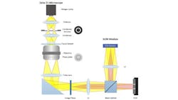
While regular microscopes use one beam of light to give the doctor a brightness map, the microscope that the research team developed uses an interferometer, which uses two beams of light—one that is kept as a reference and one that, when it goes through a sample, is modified. Comparing these two gives access to high-contrast imaging of subtle structural details in transparent samples, such as unstained tissues.
Gabriel Popescu, an Associate Professor of Electrical and Computer Engineering and the Director of the Quantitative Light Imaging Laboratory at the Beckman Institute, invented the microscope. Much of Popescu’s work to date in this area has been on prostate cancer.
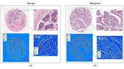
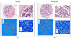
Hassaan Majeed, a researcher in Popescu's lab and one of the lead authors of the study, has been using the label-free method in targeting breast cancer cells. This high-throughput label-free imaging modality has shown imaging resolution and contrast comparable with standard histopathological imaging, while the image pixel values contain quantitative information about cellular organization.
“We measured the success of the diagnoses on our images against the gold standard,” Majeed explains. “This is a preliminary study where we are basically saying that we can resolve all the morphological markers needed by a pathologist for diagnosis. This now motivates us to move on to the next step, which is automatic tissue segmentation, using classifiers inspired by the human brain pattern recognition to perform diagnosis.”
Popescu has been in the process of developing microscopy techniques that don't require staining for the past 12 years or so. One of the major benefits of using unstained cells is that they have a longer lifespan, which means they can be studied longer. His lab has been partnering with pathologists at both Presence Covenant Medical Center (Urbana, IL) and at the University of Illinois Hospital in Chicago.
Because researchers in his lab like Majeed have shown that it is possible to obtain quantitative information from a cell, that information can be shared over a large network. That’s where the research becomes a Big Data project, using computation to even more accurately make diagnoses, prognoses, and individualize treatment.
This method compares closely with the computational advances, for example, in facial recognition, fingerprint reading, etc. By putting all the biopsies on a server (some 200 million procedures per year in the U.S. alone), doctors will have the ability to better correlate the patient’s prognosis and treatment based on correlations with a vast repository.
Popescu realizes that in order for the technique to be accepted in the community, it must be validated. The group has trained a number of pathologists on the technique, who in turn are comparing results with those of current methods for diagnosis. Popescu has also started a company called Phi Optics, which sells an interferometric imaging module that attaches to existing microscopes and incorporates this latest technology. The company was launched with funding from the National Science Foundation SBIR program as well as a seed round led by Serra Ventures out of Champaign.
Popescu adds that while researchers are scratching the surface in this quantitative phase imaging, the field is growing fast. At least 10 startups across the world are manufacturing similar optical devices for the academic environment.
Full details of the work appear in the Journal of Biomedical Optics; for more information, please visit http://dx.doi.org/10.1117/1.JBO.20.11.111210.
Follow us on Twitter, 'like' us on Facebook, connect with us on Google+, and join our group on LinkedIn


