SPECTRAL IMAGING/ONCOLOGY: New microscopy method and 3D culture enable study of breast cancer risk factors
A new imaging technology called vibrational spectral microscopy reveals subtle changes in breast tissue, promising to predict breast cancer risk and help study ways to prevent it. Developed at Purdue University (West Lafayette, IN), the technology identifies and tracks certain molecules by measuring their vibration with a laser.1 It employs a Raman spectroscopy-based technique that works at high speed, enabling measurement of changes in living tissue in real time.
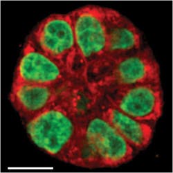
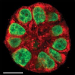
Using a "3D culture" that mimics living breast tissue, the researchers learned that a fatty acid found in some foods influences the early precancerous stage. Unlike conventional cell cultures, which are flat, the 3D cultures have the round shape of milk-producing glands and behave like real tissue, says Sophie Lelièvre, an associate professor of basic medical sciences.
The researchers are studying changes in epithelial cells, which occur where 90 percent of cancers are found. The changes in breast tissue are thought to be necessary for tumors to form, she says. "By mimicking the early stage conducive to tumors and using a new imaging tool, our goal is to be able to measure this change and then take steps to prevent it." Ji-Xin Cheng, associate professor of biomedical engineering and chemistry, adds that by monitoring the same 3D culture before and after exposure to certain risk factors, the new technique will enable detection of subtle changes in single live cells. Cheng said his lab will continue to develop a hyperspectral imaging system capable of not only imaging a specific point in a culture, but also many locations to form a point-by-point map so that tissue polarity can be directly visualized.
1. S. Yue et al., J. Biophys., 102, 5, 1215–1223 (2012).
More BioOptics World Current Issue Articles
More BioOptics World Archives Issue Articles

