NANOTECHNOLOGY/IN-VIVO IMAGING: Nanoparticle tracking analysis enables more comprehensive characterization of nanobiotechnologies
Nanoconstructs for in-vivo biomedical applications need to be in the 10–100 nm range for clearance from the kidney and reticuloendothelial system (RES), and for optical imaging and nanodrug delivery (where the nanoparticle dose administered must be determined), they also must be physiologically stable (non-aggregated). To compare plasmonic properties (the enhanced electromagnetic properties of nanoparticles), researchers further need to be able to determine the effect of different sizes and to understand the particle size distribution of similar concentrations.
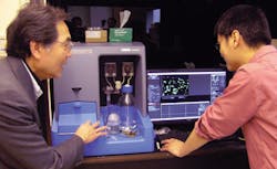
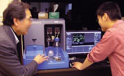
Transmission electron microscopy (TEM) and other techniques can be helpful, but have limitations. For instance, TEM allows examination of particle shape and size—but surface coating and aggregation state cannot be easily investigated using that method alone. So scientists at Duke University (Durham, NC)'s Fitzpatrick Institute for Photonics, led by Professor Tuan Vo-Dinh, have paired TEM with nanoparticle tracking analysis (NTA) for use in biosensing, imaging, and cancer therapy. The NTA-TEM combination reveals hydrodynamic size distribution and zeta potential, and allows normalization of comparison by individual particle counting.
1. Y. Hsiangkuo et al., Nanotechnol., 23, 075102 (2012).
More BioOptics World Current Issue Articles
More BioOptics World Archives Issue Articles

