GENOMICS/ATOMIC FORCE MICROSCOPY: AFM collaboration produces first in-situ view of DNA's double helix
Bart Hoogenboom uses atomic force microscopy (AFM) to achieve spatial resolution on large biomolecules in situ of about 1 nm. Hoogenboom is a lecturer with University College London (UCL) and the London Centre for Nanotechnology (LCN; a joint venture between UCL and Imperial College London), where he is also lead scientist for LCN's AFM facilities. In addition to simple visualization, AFM enables his laboratory's study of how biomolecules work.
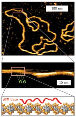
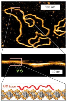
Hoogenboom's team collaborates with JPK Instruments AG (Berlin, Germany) to push instrumentation to new limits of resolution and imaging. The collaboration recently resulted in a publication in Nano Letters that reported the first visualization of the DNA double helix in water. "The resolution obtained on DNA is an example of our success in extending the capabilities of AFM instrumentation," said Hoogenboom. "Most present-day microscopes do not achieve any higher resolution on DNA than was achieved with the first AFM experiments in the early 1990s."
Hoogenboom notes that, "The distinctive feature of our recent results is the visualization of both DNA strands in the double helix. It is not new that DNA is a double helix, but about 60 years after its discovery, it is the first time that we see it in the molecule's natural, aqueous environment."

