MICROSCOPY/ CANCER DETECTION: Quantitative approach uses tissue refractive index for cellular-level cancer detection
Spatial light interference microscopy (SLIM), a label-free, automated technique that enables quantitative visualization of nanoscale structures, uses phase-contrast microscopy and holography to combine multiple light waves. Now, researchers from the Beckman Institute, Christie Clinic (Urbana, IL) and the University of Illinois at Chicago report success in using the method for cancer detection.1
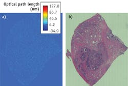
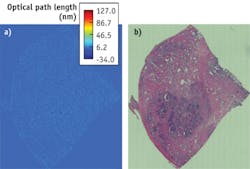
SLIM was developed by Gabriel Popescu, director of Beckman's Quantitative Light Imaging Laboratory (QLI), who also led the research—which demonstrates the value of an in-situ alternative to the limited standard histopathology approach. The scientists examined more than 1,200 biopsies, and were able to visualize unstained cells in high resolution and high contrast.
"These optical maps report on subtle, nanoscale morphological properties of tissues and cells that cannot be recovered by common stains, including hematoxylin and eosin," the researchers report. They found that cancer progression significantly alters tissue organization, which is exhibited by consistently higher refractive index variance in prostate tumors versus normal regions. The results show that the tissue refractive index reports on the nanoscale tissue architecture and, in principle, can be used as an intrinsic marker for cancer diagnosis.
Popescu says that only a small number of molecules arranged in a certain way can produce an optical signal that "something is going to happen." The team is working towards a goal of detecting cancer at the single-cell level, when the disease process is reversible. "We know that the disease starts...at the molecular level, and we think we have the proper tool to catch these early events," he notes.
A critical advantage of SLIM is that it provides quantitative, objective information. By comparison, current diagnosis methods are subjective and pathologists frequently disagree, which can lead to overtreatment.
1. Z. Wang, A. Balla, K. Tangella, and G. Popescu, J. Biomed. Opt., 16, 11 (2011).
More BioOptics World Current Issue Articles
More BioOptics World Archives Issue Articles

