MICROSCOPY/IN-VIVO CELL IMAGING: Super-resolution multifocal imaging highlights cellular detail

July 1, 2012
A hybrid approach to super-resolution microscopy called multifocal structured illumination microscopy (MSIM) has enabled researchers to see living cell structures measuring an eighth of a micrometer in size within living zebrafish larvae.

A hybrid approach to super-resolution microscopy called multifocal structured illumination microscopy (MSIM) has enabled researchers to see living cell structures measuring an eighth of a micrometer in size within living zebrafish larvae. The method combines the super-resolution capability of structured illumination microscopy (SIM) with the physical optical sectioning of confocal microscopy. It uses commercial off-the-shelf parts and open-source software, enabling integration with existing microscopes.1

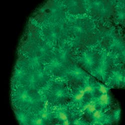
MSIM uses green fluorescent light to highlight cell structures in living fish embryos, including microtubuli—the cellular skeleton, which measure around 100 μm in length and 20 nm in diameter. (Image courtesy of NIH and KIT)

Multifocal SIM minimizes scattered light by illuminating objects only at certain spots rather than completely. Scientists can capture a series of images at variable illumination and within a few seconds—adjusting the depth of field and imaging at various depth levels. Then, they process these images on a computer to produce a 3D view. The result is sharp, illuminated detail: The method makes possible resolutions of 145 nm in the plane and 400 nm in between.

MSIM was developed by researchers from the Karlsruhe Institute of Technology (KIT; Karlsruhe, Germany), the Max Planck Institute for Polymer Research (Mainz, Germany), and the American National Institutes of Health (NIH; Bethesda, MD).

1. A. G. York, Nat. Meth., 9, 749–754 (2012).

More BioOptics World Current Issue Articles
More BioOptics World Archives Issue Articles
Sponsored Recommendations

What are Notch Filters?

Feb. 27, 2025
Notch filters are ideal for applications that require nearly complete rejection of a laser line while passing as much non-laser light as possible.

Using Optical Filters to Optimize Illumination in Fluorescence and Raman Systems

Feb. 27, 2025
Discover how Semrock products can help you get the most out of your fluorescence and Raman excitation designs, regardless of light source.

Melles Griot Optical Systems and Semrock Optical Filters for Spatial Biology

Feb. 26, 2025
Discover why a robust, high-throughput fluorescence imaging system with Semrock optical filters is key for Spatial Biology.

Understanding Practical Uses and Optimization Techniques for Fluorescence Optical Filters

Feb. 26, 2025
Learn about optical fluorescence and which optical filters to include in your instrument set up. See more with Semrock filter sets.

Voice your opinion!

To join the conversation, and become an exclusive member of Laser Focus World, create an account today!