MICROSCOPY/LIVE CELL IMAGING: Atom-shadow photo approach may advance cell biology

Life scientists are continually walking an edge: Light is needed to see, but too much can damage delicate tissues. "We can now predict how much light is needed to observe processes within cells, under optimum microscopy conditions, without crossing the threshold and destroying them," says Erik Streed, a member of the Griffith University (Brisbane, Australia) team that has photographed the shadow of a single atom for the first time, and lead author on a paper describing the work.1
According to Professor Dave Kielpinski of Griffith's Centre for Quantum Dynamics, the researchers were looking to find how few atoms are required to cast a shadow. The answer they found could have a big impact on life sciences. As reported by National Geographic, the technique could enable label-free study of DNA inside living cells by observing patterns of light absorption.
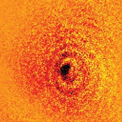
Kielpinski told BioOptics World that the team built its super-high-resolution microscope based on a phase Fresnel lens fabricated by Margit Ferstl at the Fraunhofer Institute (Berlin, Germany)—and also constructed the atom trapping apparatus, which is the bulk of the work. The apparatus uses electric fields to levitate a single atomic ion in an ultra-high vacuum chamber.
The team uses lasers "to cool down the atom and probe its optical properties. The wavelength is 369.5 nm, tuned to an atomic transition of our trapped ytterbium ion. We actually chose ytterbium ions because the lasers are easy to construct, rather than the other way around. Any atomic element would exhibit the same behavior if we used the appropriate lasers, but those lasers would generally be much harder to construct," Kielpinski said. "If we change the frequency of the light we shine on the atom by just one part in a billion, the image can no longer be seen."
"The optics setup is much like a standard microscope, except that the Fresnel lens has very small aberration, even given its large working distance (3 mm) and relatively large numerical aperture (NA = 0.65)—so we can get extremely high resolution."
1. E.W. Streed, A. Jechow, B.G. Norton, and D. Kielpinski, Nat. Comm., 3, 933, doi:10.1038/ncomms1944 (2012).