MICROSCOPY/CANCER TREATMENT: Nonlinear microscopy technique promises to reduce cancer surgeries

Nearly 40 percent of patients undergoing breast-conserving cancer surgery need subsequent surgery to remove tumor margins that are positive (cancerous) or close (suspicious). That's why oncologists and patients alike yearn for quick assessment of resected tissue during the initial operation.
A new study describes an optical nonlinear microscopy (NLM) technique that identified tumor margins with 94 percent overall accuracy on fresh, unprepared samples in situ.1 Frozen-section analysis, currently the most widely used method for intraoperative margin assessment, is time-consuming, has limited diagnostic accuracy, and produces freezing artifacts. The NLM method accomplishes rapid assessment on fresh, intact specimens without need for fixation, embedding, and sectioning required for conventional histology.
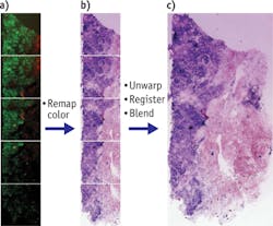
James G. Fujimoto, Elihu Thomson Professor of Electrical Engineering at Massachusetts Insitute of Technology (MIT), led researchers from MIT, Harvard Medical School (both in Cambridge, MA), and Thorlabs (Newton, NJ) in developing the technique. They used a 100 fs tunable Ti:sapphire laser at 740 nm with a 76 MHz repetition rate, and a commercially available nonlinear microscope able to image cancer-related morphological changes, such as collagen reorganization, in high resolution during surgery. Two detection channels collected two-photon fluorescence (TPF) and second-harmonic generation (SHG) signals for nuclear and stromal contrast (respectively) of the samples.
The researchers used scanning and field mosaicking to overcome limited field of view (FOV) in high-magnification imaging, and produced subcellular resolution in square-centimeter-sized specimens. They are currently redesigning the microscope to increase FOV and imaging speed, and say that mosaicking would ultimately not be required to image specimen slices. The feasibility of imaging the surface of entire specimens would ultimately depend on NLM imaging speed and specimen size, the researchers add.
The team says the technique could also be applied to lung, thyroid, and head and neck cancer surgery assessments.
1. Y. K. Tao et al., Proc. Nat. Acad. Sci., 111, 43, 15304–15309 (2014); doi:10.1073/pnas.1416955111.