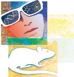
My eyes sweep across a lab rat and I can see everything. Images zoom in and out just because my brain starts thinking of looking at different scales. As my eyes pan along the length of the animal, I watch the trafficking of specific biochemicals, and I know which was which—all without labels, even without any apparent device at all.
If there were a genie in my “microscope,” this seemingly invisible technology—whatever it is—would be my first wish. But what do other biologists want from optics? I started asking around to find out.
Scientists think really big (or, rather, small) when asked: If you could get imaging to do one more thing, assuming that nothing was impossible, what advance in imaging would help you the most? “Create optical microscopes that have no limits in resolution,” says Robert LaBelle, director of marketing in the life-science research division at Leica Microsystems North America (Bannockburn, IL). Others want better resolution. Robert Doebler, director of the Marsh A. Cooper Bioengineering Laboratory at the Keck Graduate Institute (Claremont, CA), says, “To be able to achieve the submicron resolution that is achieved typically with transmission electron spectroscopy using optical fluorescence microscopy would be a huge advance.”
Scientists also wish to image intact organisms in their living environment at the highest resolution. “The inherent scattering of light in biological tissue imposes fundamental limits on the depth penetration of today’s microscopes,” says Ralf Wolleschensky, head of advanced development at Carl Zeiss MicroImaging (Jena, Germany). “An imaging system that could conquer these boundaries would truly revolutionize the industry and push science to the next level.”
Others are more specific. Marshall Montrose, chairman of the department of molecular and cellular physiology at the University of Cincinnati in Ohio, wants “whole-mouse fluorescence imaging at subcellular resolution.” Jurek Dobrucki, head of the division of cell biophysics at Jagiellonian University (Krakow, Poland), wishes for “nonphototoxic, single-molecule-fluorescence, live-cell imaging.”
Some researchers dream of more dimensions. “The living cell is four dimensional: x, y, z, and time,” says Ian Young, chairman of the department of imaging science and technology at Delft University of Technology (The Netherlands). “To understand what is going on in healthy and diseased cells, we need access to the biophysical and biochemical processes that occur in all four dimensions. It is the scales of these dimensions that make this nigh on to impossible. Spatial scales are measured in nanometers, and the temporal scale in ... choose your poison.”
It’s not just higher resolution or more dimensions that researchers want. Scientists also want better illumination. “Optical coherence tomography and microscopy have had an enormous impact,” says Richard Haskell, professor of physics at Harvey Mudd College (Claremont, CA). “But that impact would be considerably amplified if we had a cheap, stable, broadband light source with a nice Gaussian-shaped spectrum. Here’s hoping!”
Real-world expectations
Biologists also long for more-realistic advances in imaging. When asked about improvements that seem possible in the next few years, Dobrucki wishes for “very high sensitivity in fluorescence microscopy.” In fact, many biologists continue to want more from fluorescent microscopy. “I wouldn’t mind seeing more LED-based fluorescence microscopes in the near future as LEDs become more powerful, which could decrease prices, size of scopes, and maintenance time,” says Doebler.
Some advances in illumination might be closer than we think. LaBelle, for example, hopes to see “a single laser source that would cover all microscopy needs across the visible spectrum.” Other researchers hope that imaging will soon delve deeper below the surface of samples. Montrose, for example, wants to see “faster, more-sensitive, deeper penetration,” or what he calls “basically upgrades to in vivo imaging.”
It’s not just better technology that researchers want, however; they’d also like to get more for less—less money, that is. “Factor-of-two advances are nice,” says Young, “but what is really needed are factor-of-10 advances. We should be able to get 10 nm spatial resolution at a price that is one-tenth the current costs.” Still, he hopes that the near future brings better technology, too. “Acquisition times in 4-D should be on the order of seconds for chromosome-size objects,” he says.
Some of today’s developmental optics should also become more readily available soon. “A handful of new high-resolution microscopy techniques—with resolution below the diffraction-limited, ‘conventional’ microscopes—have been demonstrated in labs recently,” says Wolleschensky.
Dreams realized
As I started thinking about optical wishes for the future, I also wondered what past advances made the most difference. In asking biologists about that, I was not surprised to hear the mention of confocal microscopy—what Montrose calls “sensitive confocal imaging that lets me use living cells and tissues without turning them into dead cells and tissues.” More than one biologist also described the great value of fluorescent dyes for tagging multiple targets and better digital cameras that keep track of these labels.
Wolleschensky also provides a nice roundup of recent dreams that have come true. “Over the past decade, we have seen the development and the widespread adoption of new fluorescent proteins for biological imaging,” he says, “but what really made scientists happy was the progress in spectral-imaging hardware and unmixing software.”
As these experts reveal, the hard-working scientists focused on making these advances happen have fulfilled many wishes. Now I just want to rub a microscope, wish for my do-it-all imaging technology, and hear: “Your wish is my command!”
About the Author
Mike May
Contributing Editor, BioOptics World
Mike May writes about instrumentation design and application for BioOptics World. He earned his Ph.D. in neurobiology and behavior from Cornell University and is a member of Sigma Xi: The Scientific Research Society. He has written two books and scores of articles in the field of biomedicine.
