Medical and biological diagnosis techniques and systems are advancing at a rapid pace. Bioimaging is among those at the forefront; some experts say this technology could become the “diagnostic pillar that will lead to a lot of new breakthroughs.”
The state of the global market supports that, as the bioimaging sector hit more than $56 billion in 2020. And analysts at Research & Markets expect (conservatively) that between now and 2026, this market will grow steadily at a compound annual growth rate (CAGR) of about 10% annually. Such growth can be attributed, in part, to the “increasing prevalence of chronic medical ailments” as well as a rising elderly population with the aging “Baby Boomer” generation. This is driving the need for more advanced and efficient bioimaging systems for disease diagnosis. Bioimaging’s use for testing chemicals, toxins, and microbial materials for environmental monitoring—with the push to combat climate change—is also playing a role in this market’s growth.
The COVID-19 pandemic has also been having a direct impact on the bioimaging market, as researchers worldwide continue studying the virus and its variants to ultimately develop/enhance vaccines and other effective treatments.
Advancing the technology
Researchers are finding new ways—from more efficient and sensitive systems to more effective deployment—to get the most out of bioimaging techniques and to advance them.
A team led by the Institute of Biological and Medical Imaging (IBMI) at Helmholtz Zentrum München (Munich, Germany) has long been studying bioimaging, including with optoacoustics. A method that “relies on reading out ultrasound signals generated by light,” optoacoustic bioimaging is able to deliver a combination of high penetration depth and resolution as well as large fields of view.
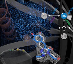
Optoacoustics relies on genetically encoded reporters and sensors, among other tools, to be effective. It also relies on reversibly photoswitchable proteins. However, traditionally, the two haven’t come together—such proteins “have yet to be used as sensors that measure the distribution of specific analytes at the nanoscale or in the tissues of live animals.” Now, in a study published in Nature Biotechnology, the IBMI team has found a way to get past that—a photoswitchable calcium ion sensor they’ve developed (see Fig. 1), based on a 5G genetically encoded calcium indicator (GCaMP5G). It can be switched with 405/488 nm light and can describe the molecular mechanisms at the structural level.1
According to the researchers, “the light switchable signal enables us to visualize small numbers of cells against a strong background of other signals by making the label blink. The ability to visualize few cells in a live organism is important because many biological phenomena, especially in the immune system, rely on a small number of cells.”
The IBMI team is working to eventually be able “to track single-labeled cells in a living organism and visualize their function” and thus better understand areas such as the immune system and tumor development.
Researchers from the Beijing National Laboratory for Molecular Sciences (China) are forwarding the use of fluorescence in bioimaging. With emission wavelengths in the near-infrared (near-IR) range (700–1400 nm), fluorescent dyes allow tissue imaging from deep within a sample. While dyes in the near-IR-II range (1000–1700 nm) could also boost bioimaging, they are hindered in their lack of brightness.
Now, the Beijing team has developed new dyes that strongly fluoresce in the near-IR-II. The new xanthene-based family of dyes has exhibited “the best performance with fluorescence emission at 1210 nm and high brightness.”
In their study—published in the Journal of the American Chemical Society—the researchers demonstrated their findings in the blood circulation of mice.2 After injecting the dye, the team observed the animal’s circulatory system lighting up as the dye progressed through its body. “The dye was bright enough that clear images could be obtained with exposure times as low as 5 ms,” according to the study, which allowed the researchers to calculate blood-flow velocities and achieve “spatial resolution that was good enough to differentiate between closely spaced femoral arteries and veins.”
The researchers say this is “an effective tool for high spatial and temporal resolution bioimaging.”
Researchers from Okinawa Institute of Science and Technology Graduate University (OIST), in conjunction with a team from Kyushu University, both in Japan, are also taking steps toward advancing bioimaging. They have been able to generate a glow-in-the-dark light using organic materials, rather than inorganic crystals, which are needed to ensure a high level of bioimaging system performance. The inorganic route used currently requires the implementation of rare-earth metals that are not easily found, as well as fabrication temperatures of over 1800°F.
The organic materials are more readily available, making them ideal. The OIST and Kyushu researchers say bioimaging is a strong application candidate for their organic-based light and could produce a myriad of benefits for the health sciences realm.
“Not only are organic materials much more available and easier to work with than inorganic materials, but they are also soluble, which has the potential to diversify and expand the use of glow-in-the-dark objects” for applications including bioimaging, according to researcher Chihaya Adachi, a professor and director of the Center for Organic Photonics and Electronics Research at Kyushu University.
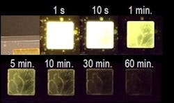
In a 2017 study published in Nature, Adachi and Ryota Kabe, a professor who leads the Organic Optoelectronics Unit at OIST, demonstrated (for the first time) that two organic materials could create a glow-in-the-dark effect.3 At that time, however, its performance with two materials was about 100 times weaker than systems that employed inorganic materials. Now, these researchers have found that advancing to three organic materials as well as a change in molecules boost the strength (see Fig. 2). The team notes “a tenfold improvement from the previous work.”“With organics, we have a great opportunity to reduce the cost of glow-in-the-dark materials, Adachi says, adding that exploiting the versatility of organic materials could lead to technologies such as “bio-compatible probes for medical imaging."
Future potential
As the bioimaging market is expected to “witness stunning growth by 2028,” according to A2Z Market Research, the pool of those exploring new technologies, advancements, and techniques is growing. Not only are scientists and other researchers involved, but now outside players as well.
Investors and funding initiatives are now understanding the benefits of more advanced bioimaging systems—in the U.S. and beyond. Among those acknowledging the need is the Chan Zuckerberg Initiative (CZI). The organization recently awarded $5 million “to advance bioimaging technologies, increase access to these tools, and build capacity for biomedical researchers.”
“Expanding imaging capacity for biomedical researchers requires advancing imaging software and hardware, expanding access to shared tools and resources, and building capacity for imaging scientists and organizations to advance research in their home countries,” says Stephani Otte, Imaging Program Officer for CZI.
Specifically, $1 million of the funds will support plugin projects for napari—“a community-built, Python-based, open-source tool designed for browsing, annotating, and analyzing large multidimensional images, utilized by practitioners, biologists, and other scientists.” CZI notes that this is essential to biomedical science and its advancement.
The remaining funds will support Expanding Global Access to Bioimaging projects that will increase access to imaging instrumentation and expertise for biomedical researchers in Africa, Latin America and the Caribbean, and former Soviet countries. These projects, according to CZI, will “expand access to imaging expertise, technologies, and capacity building for researchers. Teams foster collaborations between imaging scientists and biomedical researchers and increase representation of regional imaging scientists in the global community.”
“To cure, prevent, or manage all diseases,” says Vladimir Ghukasyan, Imaging Community Program lead for CZI, “we need to make sure that scientists around the world have access to top technology and expertise.”
About the Author
Justine Murphy
Multimedia Director, Digital Infrastructure
Justine Murphy is the multimedia director for Endeavor Business Media's Digital Infrastructure Group. She is a multiple award-winning writer and editor with more 20 years of experience in newspaper publishing as well as public relations, marketing, and communications. For nearly 10 years, she has covered all facets of the optics and photonics industry as an editor, writer, web news anchor, and podcast host for an internationally reaching magazine publishing company. Her work has earned accolades from the New England Press Association as well as the SIIA/Jesse H. Neal Awards. She received a B.A. from the Massachusetts College of Liberal Arts.


