Microscopy breakthrough takes biological research to new depths
Since the very first observation of fluorescence was reported by Sir Frederik William Herschel in 1845, and its first actual description presented by British scientist Sir George G. Stokes in 1852, its use in microscopy has played an extremely important role in biological discovery. Scientists can see and analyze cells; from the results, they’ve been able to discover organisms and previously unknown life, as well as study and ultimately combat disease. Fluorescence microscopy has allowed scientists and engineers to make some of the most significant biological finds to date.
However, despite its effectiveness, the method has had limitations since its earliest use—shallow penetration depths into samples and cells’ sensitivity to phototoxicity, among them. And while it has evolved exponentially over the past century, some restraints have remained.
“Mammalian tissues are naturally opaque, which hinders optical imaging techniques from attaining high-resolution imagery,” says Quanyu Zhou, a researcher at the Institute for Biomedical Engineering and Institute of Pharmacology and Toxicology at the University of Zurich and the Institute for Biomedical Engineering at ETH Zurich, both in Switzerland, and a member of Daniel Razansky’s Lab.
To access deep brain regions, conventional optical microscopy techniques opt for highly invasive surgical methods. Often used to image molecular and cellular details of the brain in animal models of various diseases, fluorescence microscopy has been limited to small volumes and highly invasive procedures such as craniotomy due to intense light scattering by the skin and skull.
According to a study led by Zhou and Razansky, a professor of biomedical imaging, the effective penetration depth and resolving capacity provided by fluorescence microscopy have been limited, thanks to intense light scattering in living tissues.1 They note that while shortwave-infrared (SWIR) cameras and contrast agents featuring fluorescence emission in the second near-infrared (NIR-II) window have expanded the achievable penetration, “the effective spatial resolution progressively deteriorates with depth due to photon diffusion.”
“The penetration depth of optical microscopy is still fundamentally limited by the so-called ballistic regime (typically <1 mm from the surface),” according to Zhou and Razansky, adding that “enabling high-resolution optical observations in deep tissue represents a long-standing goal in the biomedical imaging field.”
Their team is advancing toward this goal with a new technique—diffuse optical localization imaging (DOLI)—that allows microscopic fluorescence imaging at 4X the depth limit imposed by light diffusion. This is making it a promising platform for studying neural activity, as well as microcirculation, neurovascular coupling, and neurodegeneration. According to their research, the method is based on “localization of flowing microdroplets encapsulating lead sulfide (PbS)-based quantum dots in a sequence of epifluorescence images acquired in the NIR-II spectral window.”
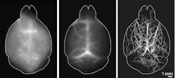
Specifically, the Zurich team’s DOLI technique takes advantage of the NIR-II spectral window spanning from 1000 to 1700 nm. This exhibits less scattering and can effectively reduce background autofluorescence signals (see figure). “By localizing a sparsely labeled contrast agent within the NIR-II window,” Razansky says, “DOLI enables superresolution imaging through a diffuse medium, such as the murine brain, noninvasively.”
Razansky’s lab specializes in development of new multiscale functional and molecular imaging techniques, with a major focus on noninvasive brain imaging. According to Razansky, right now most murine brain studies with optical microscopy are limited to small brain regions and rely on highly invasive procedures.
“The introduction of highly efficient [SWIR] cameras and bright quantum dots in the NIR-II spectral window enabled us to image deep brain regions noninvasively, while the spatial resolution was further improved by introducing on the localization concept,” he says.
The group’s goal in their recent research has been to image even deeper into the brain, for an even better and more comprehensive study of it. Visualizing biological processes deep in the intact living brain is crucial for understanding both its cognitive functions and neurodegenerative diseases such as Alzheimer’s and Parkinson’s. The researchers say that with their new DOLI method, they have been able to clearly visualize the microvasculature and blood circulation deep in the mouse brain entirely noninvasively for the first time. This ultimately enables a closer look at a living, functioning brain.
Additionally, the study has found that the DOLI technique “further enables retrieving depth information from planar fluorescence image recordings by exploiting the localized spot size.” It operates in “a resolution-depth regime previously inaccessible with optical methods, thus massively enhancing the applicability of fluorescence-based imaging techniques.”
“DOLI’s superb resolution for deep-tissue optical observations can provide functional insights into the brain,” Zhou says. “We believe with the collective efforts on more efficient [SWIR] cameras, as well as novel contrast agents exhibiting strong fluorescence responses in the NIR-II window, many unknown questions in these fields can be answered in the near future.”
REFERENCE
1. Q. Zhou, Z. Chen, J. Robin, X.-L. Deán-Ben, and D. Razansky, Optica, 8, 6, 796–803 (2021).
About the Author
Justine Murphy
Multimedia Director, Digital Infrastructure
Justine Murphy is the multimedia director for Endeavor Business Media's Digital Infrastructure Group. She is a multiple award-winning writer and editor with more 20 years of experience in newspaper publishing as well as public relations, marketing, and communications. For nearly 10 years, she has covered all facets of the optics and photonics industry as an editor, writer, web news anchor, and podcast host for an internationally reaching magazine publishing company. Her work has earned accolades from the New England Press Association as well as the SIIA/Jesse H. Neal Awards. She received a B.A. from the Massachusetts College of Liberal Arts.

