Printable biosensor could be invaluable tool for surgeons
Researchers at Purdue University (West Lafayette, IN), in conjunction with a team at Los Alamos National Laboratory (LANL; Los Alamos, NM), are aiming to assist medical professionals with a new 3D-printable biosensor that allows the simultaneous recording and imaging of tissues and organs during surgical procedures.
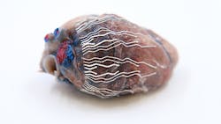
Developed by Chi Hwan Lee, an assistant professor of Biomedical Engineering in Purdue’s Weldon School of Biomedical Engineering and assistant professor of mechanical engineering, the biosensor is ultrasoft, thin, and stretchable, and capable of “seamlessly interfacing with the curvilinear surface of organs,” namely the heart. The concurrent recording and imaging ability can help surgeons to accurately indicate the origin of disease conditions; according to Lee, conventional methods have proven difficult “because other sensors used for recording typically interrupt the imaging process.”
Bio-inks were used in the biosensor prototype’s development, which are softer than tissue and can stretch without sensor degradation. Additionally, they can naturally adhere to wet surfaces of organs. Several prototypes of the biosensor—of varying sizes, shapes, and configurations—have been developed by the researchers, each tested in the hearts of pigs and mice in vivo. The researchers have also been studying the “biocompatibility and anti-biofouling properties of the biosensors,” along with their effects on cardiac function; no significant adverse effects have been reported.
The new biosensor could be further developed for use in other organs in the body, including the stomach, according to the researchers. Reference: B. Kim et al., Nat. Commun. (2021); doi.org/10.1038/s41467-021-23959-3.
About the Author

Justine Murphy
Multimedia Director, Digital Infrastructure
Justine Murphy is the multimedia director for Endeavor Business Media's Digital Infrastructure Group. She is a multiple award-winning writer and editor with more 20 years of experience in newspaper publishing as well as public relations, marketing, and communications. For nearly 10 years, she has covered all facets of the optics and photonics industry as an editor, writer, web news anchor, and podcast host for an internationally reaching magazine publishing company. Her work has earned accolades from the New England Press Association as well as the SIIA/Jesse H. Neal Awards. She received a B.A. from the Massachusetts College of Liberal Arts.