780-nm photobiomodulation advances peripheral nerve repair toward a device-ready clinical platform
Each year, more than four million people worldwide sustain a peripheral nerve injury, with falls and mechanical trauma the leading causes.1 Across the seven largest healthcare markets, close to 4 million new cases were recorded in 2023, driving nearly 1.7 million nerve-related surgeries.2
Surgical repair (autografts and synthetic nerve conduits) remains the standard of care, but outcomes have plateaued; even the autograft “gold standard” leaves many patients with incomplete recovery across long nerve gaps.3 No drug is FDA-approved to accelerate axonal regeneration after nerve injury,4 opening space for photonics-based solutions that stimulate regeneration noninvasively.
A photonics solution
Photobiomodulation (PBM) uses low-power light in the red and near-infrared range to stimulate biological activity without thermal damage. Among wavelengths studied for peripheral nerve repair, 780 nm has emerged during four decades of experimental work as the wavelength that most reliably promotes nerve regeneration,5 reflecting near-optimal tissue penetration and absorption by mitochondrial cytochrome-c oxidase.
When cytochrome-c oxidase absorbs 780-nm light, mitochondrial membrane potential and adenosine triphosphate (ATP) synthesis increase, followed by changes in reactive oxygen species and calcium signaling that alter membrane permeability and enzyme activity.6 In the injured peripheral nerve, these events translate into enhanced Schwann cell activity supporting remyelination, sustained axonal sprouting across the injury site, and preserved neuromuscular junction and acetylcholine receptor density in denervated muscle, delaying the atrophy that limits functional recovery.
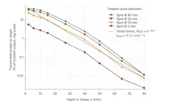
Delivery, not biology, is the limiting engineering variable. In vivo measurements through human skin and arm muscle with a 780 ± 2-nm continuous-wave (CW) diode laser at 250 mW show near-exponential attenuation with depth, and a strong dependence on treated-zone diameter (see Fig. 1). For a 40-mm zone, 97 mW reaches 2-mm depth (38.8% of output), 58 mW at 15 mm, and 6 mW at 40 mm; narrowing the zone to 2 mm drops transmitted power to 14 mW at 2-mm depth. Spot geometry and collimation therefore govern whether a deep nerve trunk receives a therapeutic dose, which is why the platform indexes dose to anatomical target rather than to applicator output alone.
Clinical translation was achieved in a double-blind, placebo-controlled randomized trial: 780-nm CW laser at 250 mW, applied transcutaneously for three hours daily over the injured nerve (450 J/mm2) and two hours over the corresponding spinal cord segments (300 J/mm2) for 21 consecutive days, produced statistically significant motor recovery in patients with long-standing incomplete peripheral nerve injuries (p = 0.0001).7 The WALT 2026 position paper on PBM dosimetry now provides a framework for standardizing these parameters for industrial deployment.8
Four decades of evidence
The clinical case for 780-nm PBM rests on a coherent body of work built over four decades, reviewed in Rochkind's 2017 summary: Mechanism, intraoperative use, reconstruction with guiding tubes, a triple-target concept, and the randomized placebo-controlled trial.
In rat models, 780-nm irradiation produces a consistent set of effects: An immediate and sustained protective effect on injured-nerve function, reduced scar-tissue formation, prevention of retrograde motor-neuron degeneration in the spinal cord, and increased axonal growth and myelination. The first clinical use was intraoperative 780-nm irradiation applied immediately after direct nerve repair. A 2007 double-blind study then combined 780-nm phototherapy with polyglycolic-acid neurotube reconstruction, showing accelerated axonal growth and improved regeneration three months after surgery.9
A 2016 rat study tested simultaneous 780-nm PBM applied to three sites: Nerve, spinal cord, and denervated muscle. Long irradiation nearly doubled large-diameter neuronal fibers and significantly reduced muscle atrophy (8.06% ± 1.23% vs. 24.44% ± 7.26%; p < 0.05)—the direct precursor to the workstation's three-outlet design, one channel per anatomical target.10
A device: Three generations of engineering
The validated clinical protocol was implemented in three successive device generations, each built for a different clinical workflow while preserving the same core photonic parameters. The first- and second-generation systems shared the same clinical operating point: A near-infrared diode-laser controller feeding a collimated head mounted on an articulating arm above the operating table, used both intraoperatively from the 1990s and for the transcutaneous ambulatory protocol of the randomized trial.
The third-generation commercial workstation now under development builds the same 780-nm CW source, transcutaneous delivery, and 21-day protocol into a mobile pantograph-arm platform with an on-cart touchscreen console, three independent output channels, a body-schematic dosing interface, and an emergency-stop safety loop (see Fig. 2).
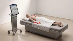
Each channel drives a separate collimated applicator, allowing nerve, spinal cord, and muscle targets to be treated in one session with channel-specific exposure times. Because transmitted power falls by more than an order of magnitude between 15- and 40-mm depth, the console derives per-channel dose from the selected anatomical site and applicator aperture rather than from a single global setting—the reproducibility gap left by existing commercial PBM devices.
Practical design targets follow directly from the clinical protocol: A stable 780-nm center wavelength with drift below 1 nm across the treatment session, 250-mW CW wave output held within a few percent over multi-hour exposures, thermal management sufficient to keep skin-surface temperature rise negligible, and per-channel optical monitoring so that an aperture blocked by patient movement is detected rather than silently reducing delivered dose.
From bench to market
In the U.S., PBM devices are regulated by the Federal Drug Administration (FDA) as Class II devices, cleared through the 510(k) pathway on the basis of substantial equivalence; in the European Union, the device falls under Regulation (EU) 2017/745 as Class IIa, requiring notified-body audit and CE marking. Neither pathway requires a full Premarket Approval, and the completed randomized trial exceeds the clinical evidence typically expected for a Class II submission. The engineering work that remains is verification rather than discovery: Wavelength and power stability during the 21-day protocol, IEC 60825-1 laser-class documentation, IEC 60601 electrical and usability compliance, and calibration traceability for each output channel.
Independent analyses place the global peripheral nerve injury market at USD $2.98B in 2026, forecast to reach USD $4.30B by 2031 at a 7.6% CAGR,11 with the seven major markets valued at ~USD $2.4B in 2023. In the U.S. alone, the market is projected to grow from USD $501M (2025) to USD $1.05B (2035).12 Established PBM manufacturers have concentrated on pain management, wound healing, and dermatology, leaving the peripheral nerve segment without a dedicated commercial device. A validated 780-nm platform enters an underserved indication as first mover, and a peripheral-nerve clearance would serve as a predicate for diabetic neuropathy, post-surgical recovery, and compression neuropathy.
Call for industrial partnership
Four decades of work have converged on a defined operating point: 780-nm, 250-mW, transcutaneous multisite delivery validated in a randomized placebo-controlled trial and now built into a third-generation clinical workstation. The remaining work is industrial, not scientific.
A partnering organization would inherit a translation-ready package: A validated wavelength and dosimetry, a published clinical protocol, a device design refined through three generations of clinical use, and a peer-reviewed evidence base spanning mechanism, preclinical models, and a placebo-controlled randomized trial. The authors are open to licensing the workstation technology and its intellectual property, codevelopment, manufacturing partnerships with medtech and photonics OEMs, and regional distribution agreements.
Enquiries are welcome from medtech OEMs, photonics manufacturers, and rehabilitation device companies. Please contact the authors to review the technical dossier, regulatory pathway, and evidence base.
REFERENCES
1. S. Shi et al., Front. Neurol., 16, 1669662, 1990–2021 (2026).
2. See www.delveinsight.com/report-store/peripheral-nerve-injuries-market.
3. S. Rochkind, A. Goldlust, and M Almog, Brain Spine, 6, 106115 (2026).
4. S. J. Hasani et al., Biochem. Biophys. Rep., 45, 102514 (2026).
5. S. Rochkind, Photomed. Laser Surg., 35, 11, 604–615 (2017).
6. T. I. Karu and S. F. Kolyakov, Photomed. Laser Surg., 23, 4, 355–361 (2005).
7. S. Rochkind, V. Drory, M. Alon, M. Nissan, and G. E. Ouaknine, Photomed. Laser Surg., 25, 5, 436–442 (2007).
8. R. Hanna et al., J. Clin. Med., 15, 3, 1304 (2026).
9. S. Rochkind et al., Photomed. Laser Surg., 25, 3, 137–143 (2007).
10. M. M. Mandelbaum-Livnat et al., Photomed. Laser Surg., 34, 12, 638–645 (2016).
11. See www.mordorintelligence.com/industry-reports/peripheral-nerve-injuries-market.
12. See www.novaoneadvisor.com/report/us-peripheral-nerve-injuries-market.
About the Author
 Shimon Rochkind
Shimon Rochkind, MD, Ph.D., is an associate professor at Tel Aviv University and head of the Microsurgical Center for Peripheral Nerve Reconstruction at Assuta Medical Center in Tel Aviv, Israel. He holds an academic affiliation with Tel Aviv University and specializes in neurosurgery and microsurgery, with particular expertise in peripheral nerve reconstruction, brachial plexus surgery, pediatric neurosurgery, and regenerative laser-based and bioengineering technologies. Rochkind earned his Ph.D. at Bar-Ilan University and completed clinical fellowships in pediatric neurosurgery in Toronto, Canada; brachial plexus and peripheral nerve surgery in Cape Town, South Africa; and brachial plexus reconstruction in Ogori, Japan. He is a former president of the Sunderland Society and of the World Association for Photobiomodulation Therapy (WALT), and serves on the EANS Committee of Peripheral Nerve Surgery of the European Association of Neurosurgical Societies. Rochkind has authored more than 140 peer-reviewed articles, review papers, case reports, and book chapters, and holds 11 international patents in laser technology and biotechnology; his research has been funded by the EU FP7 program, Teva Pharmaceutical Industries, Johnson & Johnson, Baxter International, and other national and international grants.
 Igal Koifman
Igal Koifman, BSc, MBA, is a photonics and medical-device engineer based in Israel, with more than two decades of experience in laser system development. He is a co-inventor of 13 patents spanning medical laser therapy systems, fiber-optic delivery and diagnostics, diode-laser imaging heads, and augmented-reality display systems, filed with Lumenis Ltd., Creo IL Ltd., and Yeda Research and Development Co. Ltd. (Weizmann Institute of Science). His work on handheld laser treatment platforms, cooling architectures for laser handpieces, optical-fiber integrity monitoring, and fiber-to-tissue distance estimation addresses the delivery, dosimetry, and safety problems central to clinical laser devices. He is codeveloper of the third-generation 780-nm peripheral nerve photobiomodulation workstation described in this article.