Biohybrid image sensor array processes light like retinas
A retina-inspired biohybrid image sensor array (BIOPIX) created by a team of researchers led by Thomas M. Brown, a professor of organic and biological electronic engineering at Tor Vergata University of Rome in Italy, responds to light in a way remarkably similar to the photoreceptor cells of a retina—in terms of speed and how it senses color (see video).
The team’s “retina emulator” combines organic electronics with biological liquids to convert light into electrical signals in a manner akin to a retina’s rods and cones, and generates real-time images on a display (which is believed to be the first demonstration of its kind).
“Years ago at a Materials Research Society meeting, I was deeply impressed on several levels—scientifically and in terms of innovation and the potential to improve the quality of life—by a demonstration of a man who was blind for many years receiving a silicon/metal retinal implant,” says Brown. “Afterward, he was able to identify and pick up a white cup placed against a black background. It was a profound moment for me.”
At the time, Brown was researching organic semiconductors for photovoltaics, which other researchers had shown worked for the same applications and offered flexibility and biocompatibility. It led to a collaboration with Ebin Joseph, a postdoctoral researcher, as well as Antonella Camaioni, a professor of histology in the school of medicine at Tor Vergata University of Rome, an interdisciplinary team of 15, and funding agencies interested in creating a retina emulator.
How do you build a retina emulator?
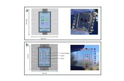
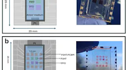
The team’s array design combines 12 pixels to mimic rod-like responses (night vision) with a central 2 × 2 array (think matrices) to simulate cone-like dichromic sensitivity (color vision in most mammals).
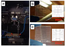
To get started, the researchers print different semiconducting conjugated polymers over transparent laser-patterned microelectrodes that absorb light with similar spectral shapes to the cone and rod photoreceptor cells in mice.
“On the same platform, we integrated a reference electrode with a platinum nanoparticle surface that can exchange charges with ions, and created a small chamber that we filled with a water-based physiological medium resembling the physiological conditions found within live retinal tissues,” explains Brown. “This electrolytic medium, by immersion, connects all of these components together electrically, which enables the device array to become optoelectronically active.”
Each polymer responds preferentially to a specific spectral region. “In our device architecture, P3HT (a p-type semiconducting polymer) shows sensitivity to green light (similar to the medium wavelength M-cone of a mouse); PFO (a wide-bandgap blue-light-emitting polymer) responds primarily to ultraviolet wavelengths (similar to the short wavelength S-cone of a mouse); and P3HT:PCBM has a similar absorption profile to P3HT but shifted toward the blue (more similar to the rod cells of a mouse),” Brown adds.
Biocompatibility of the technology platform and its materials was confirmed through in vitro testing of culturing primary human mesenchymal stromal cells over its surface. “Its good biocompatibility highlights the potential for using the electrode and semiconducting materials we used here in future biomedical applications,” says Brown.
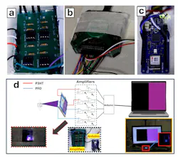
Once light is absorbed by the polymer pixels of the array, an electrical signal is created that’s conducted through the water-based physiological medium to the reference electrodes. “The electrical signals coming out of our array resemble in shape, as well as characteristic times, those found within mouse photoreceptor cells,” Brown points out. “And the electrical signals generated by each pixel are then processed through a dedicated electronic readout system, which allows us to create pixelated images in real time—demonstrating color sensitivity (through the artificial cone pixels) and grayscale contrast (through the artificial rod pixels). We believe this is the first demonstration of imaging on a display in real time through a liquid/solid biohybrid sensor array.”
Electronic readout system/display
While the team didn’t use computational simulations for their work, to convert optical signals captured by BIOPIX into direct-to-display pixelated images (with a dichromatic color palette) they developed a dedicated electronic readout system—including customized software designed to resolve the dichromatic vision type and retina-like ionic temporal dynamics of mice retinas.
“Future studies could benefit from optical and electrochemical simulations to further optimize polymer absorption profiles, pixel geometry, and charge transport dynamics at the polymer-electrolyte interface,” says Brown.
Sensor signals at timescales of retinas
While designed for this, “one of the most satisfying moments of our work was when we finally observed the device array produce distinct electrical responses to different colored light,” says Brown. “The most exciting moment was seeing the light incident on the sensor being converted into pixelated images on a screen in real time—which was a first.”
A big moment for the team was when they discovered “detectable signals from our sensors were for light pulses down to 20- to 50-millisecond durations,” Brown adds. “These are the typical ‘slow’ timescales found within real living retinas—solid-state image sensors have orders of magnitude faster response times.”
Hurdles along the way
The team faced a few challenges during their device’s development. The first was to establish a simple and reliable fabrication technique to create polymer pixel arrays. “We developed a stencil-printing approach to significantly simplify the fabrication process and make pixelation faster and more reproducible,” says Brown.
Another challenge was small photocurrents generated by the hybrid organic semiconductor/liquid photodetectors, which were often in the nanoampere range or even below. “Detecting such weak signals requires careful signal amplification and noise reduction—particularly when converting the sensor output into direct-to-display still images using a customized analog-to-digital system designed by our team,” Brown says. “Device stability was also an important consideration. Ensuring stable operation within the chamber filled with a water-based physiological medium required careful selection of materials and thoughtful device design to maintain reliable performance.”
An introduction to the BIOPIX color image sensor array technology. Credit: Tor Vergata University of Rome
Proof of concept
The team’s BIOPIX retina emulator platform can be used to explore new photoabsorbing artificial photoreceptor materials and physiological media prior to retinal implantation or injection, as well as to evaluate their performance within different environmental conditions.
It can also help explain differences in image sensing operating in a fully solid-state mode vs. at the interface between biological (liquid) and semiconducting (solid) matter within a field where biology and technology are coming together to enable new possibilities. “Understanding the underlying biophysics of light, semiconductors, and biological matter can help in the quest to restore vision to those who’ve lost it through degenerative retinal diseases or help to improve function for the visually impaired,” says Brown.
This work is a “proof of concept of a biohybrid pixel sensor array,” says Brown. “During the next five years, we’d like to improve sensitivity and signal amplification while reducing the pixel-level crosstalk, as well as increase pixel density for higher-resolution sensing. We’d also like to test its long-term stability within biological environments, design new materials for future implantation/injection, explore integration with living cells, and use it to excite and sense living cells. Medical research will follow.”
FURTHER READING
E. Joseph et al., Adv. Mater. Technol., 11, 2, e01461 (2026); https://doi.org/10.1002/admt.202501461.
Related Webinar
About the Author

Sally Cole Johnson
Editor in Chief
Sally Cole Johnson is Laser Focus World’s editor in chief, and she has more than 25 years’ experience as a science and technology journalist. She specializes in physics and semiconductors, and wrote for the American Institute of Physics for more than 15 years, and also covered theoretical physics and neuroscience for the Kavli Foundation, and complexity for the Santa Fe Institute. Johnson has also written extensively about military embedded systems, high-performance computing, software-defined networks, and infosec. She is a member of the National Association of Science Writers (since 2001).
When she isn’t writing about optics, photonics, or quantum advances, you can find her outside in northern NH in the garden with birds landing in her hand or heading for the mountains with her bike, skis, or crampons and ice axe.