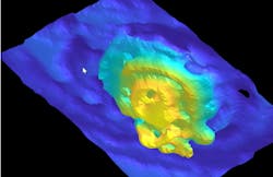
Terahertz near-field imaging approach goes deep into the ear
A team at Waseda University (Shinjuku, Japan)—working with researchers from the Institute of Laser Engineering at Japan’s Osaka University, Kobe University (Hyogo, Japan), the Georgia Institute of Technology (Atlanta, GA), and the Research Institute for Interdisciplinary Science at Okayama University (Okayama, Japan)—has developed a 3D terahertz near-field imaging technique to examine the cochlea, a spiral-shaped organ within the inner ear responsible for hearing; it converts sound waves into neural signals (see video).
Given the delicate cochlea’s position within the ear, it’s difficult to see with conventional imaging methods, which can only get down to millimeter focus sizes.
Systems involving terahertz radiation are ideal for biological imaging because they emit low energy and don’t harm tissues. They also create less scattering than near-infrared (NIR) and visible light, and can image through bone but are also sensitive to changes in hydration and cellular structure.
“It’s because the cochlea is surrounded by bone,” says research team leader Kazunori Serita, an associate professor at Waseda. “Light can’t get through it, and x-rays come with radiation risks for the organs being exposed. With conventional methods, there just wasn’t a safe way to see inside.”
And seeing inside is crucial for making a proper diagnosis.
Listen up!
In their work involving a mouse model, the team used a terahertz point source (a terahertz wave source) with a diameter of merely 20 µm to shine light directly onto the sample, which allows detection of terahertz waves that reflect from the inside the ear. By detecting those terahertz waves, the team reconstructed detailed 3D images and could view the sample in cross-sectional slices—similar to a CT scan.
“Since our terahertz point source is on a micron scale, it allows us to see the cochlea’s tiny internal structures in detail,” Serita says.
The team originally built the imaging system just to get high-resolution 2D images of the cochlea, which was successful. At the same time, they also recorded time-domain waveforms. “Just by chance, we spotted a tiny reflected signal in one of them,” Serita says. They then wondered if by syncing the imaging with those tiny reflections, perhaps they could go further and obtain a 3D view of the inside.
“We got really good resolution!” Serita says, noting there have been several previous reports on 3D terahertz imaging, but the spatial resolution wasn’t great. The conventional approaches make imaging such small samples out of the question.
This new method doesn’t rely on a lens, Serita explains. Instead, they used the tiny terahertz point source that forms when terahertz waves are generated from light inside the nonlinear optical crystal.
“Since this point source is on a micron scale, it allows us to measure much smaller samples like cells with terahertz waves,” Serita says.
Imaging the future
So far, the team has tested their terahertz imaging technique on extracted and dried mouse cochleae to evaluate its effectiveness. Their next step? Demonstrate the method’s feasibility on actual cochleae in a more realistic biological environment.
“Since the cochlea is located deep inside the ear and filled with lymphatic fluid (water), several improvements are needed,” Serita says. “We need to miniaturize the system so it can be inserted through the ear canal and develop a stronger terahertz source to penetrate deeper structures.”
The team’s ultimate goal is to make it possible to diagnose hearing loss and other ear conditions in vivo. Terahertz waves could be one of the ways to do that.
“Our work could lead to a new diagnostic method for ear diseases that have been difficult to diagnose until now. It has the potential to enable onsite diagnosis of conditions like sensorineural hearing loss and other ear disorders,” Serita says. “In the future, this technology may also help with early detection of hearing impairments and allow for earlier treatment and better outcomes for patients.”
FURTHER READING
L. Zheng et al., Optica, 12, 4 (2025); https://doi.org/10.1364/optica.543436.
About the Author
Justine Murphy
Multimedia Director, Digital Infrastructure
Justine Murphy is the multimedia director for Endeavor Business Media's Digital Infrastructure Group. She is a multiple award-winning writer and editor with more 20 years of experience in newspaper publishing as well as public relations, marketing, and communications. For nearly 10 years, she has covered all facets of the optics and photonics industry as an editor, writer, web news anchor, and podcast host for an internationally reaching magazine publishing company. Her work has earned accolades from the New England Press Association as well as the SIIA/Jesse H. Neal Awards. She received a B.A. from the Massachusetts College of Liberal Arts.


