Enhance early detection of melanoma with liquid lenses
Melanoma, a deadly form of skin cancer, poses a significant global health concern. Early detection is crucial for successful treatment, but it remains challenging due to the complexity of skin lesions and limitations of imaging technologies. Fortunately, advancements in optics and imaging are revolutionizing melanoma detection.
Within the context of melanoma detection, optics focuses on the development and refinement of tools and technologies that manipulate light for better visualization of skin lesions. It can include:
Dermatoscopes. Handheld devices that use polarized light to enhance visualization of skin structures.
Laser-based systems. Tools that use specific wavelengths of light to analyze skin tissue properties.
Optical coherence tomography (OCT). A noninvasive imaging technique that uses light waves to take cross-sectional pictures of skin layers to help evaluate lesion depth and structure.
Imaging involves capturing detailed images of skin lesions and using various techniques to enhance, analyze, and interpret these images for diagnostic purposes. This includes:
Digital dermoscopy. High-resolution digital images of skin lesions are taken with a dermatoscope, which can be analyzed over time for changes.
Reflectance confocal microscopy (RCM). A technique that provides cellular-level resolution images of the skin, and enables detailed examination of lesion morphology.
Multispectral imaging. Multiple wavelengths of light capture images that highlight different aspects of skin lesions, which helps to differentiate between benign and malignant lesions.
Optics and imaging technologies are often integrated to provide comprehensive tools for melanoma detection. For example, a dermatoscope (an optical device) can be used to capture high-resolution images (imaging) that are then analyzed using software to identify potential melanoma.
Recent advancements often combine optical principles with advanced imaging techniques to improve diagnostic accuracy. Some modern systems use multispectral imaging combined with machine learning algorithms to analyze images more effectively.
Understanding melanoma
Melanoma, a type of skin cancer, arises when melanocytes—the pigment-producing cells in the skin—undergo malignant transformation. It can manifest as unusual moles, lesions, or discolored patches on the skin. While early-stage melanoma is typically curable, advanced melanoma has the potential to spread to other parts of the body and it leads to a poor prognosis and lower survival rates.
Challenges in detection
Early detection of melanoma demands keen observation and sophisticated imaging tools, because traditional visual inspection and dermoscopy often struggle to reliably differentiate benign from malignant lesions. Factors such as lesion depth and irregular borders can further complicate accurate diagnosis.
This is where the Intelligent Total Body Scanner for Early Detection of Melanoma (iToBoS) project, funded by the European Union’s Horizon Europe program, comes into play. Through iToBoS, multiple partners are developing an artificial intelligence (AI) platform that helps detect melanoma. The platform includes a total body scanner and computer-aided diagnosis tool that integrate patient-specific data like age, sex, anatomical skin lesion locations, the number and size of moles, and more. Based on a prototype developed by three project partners and powered by Optotune’s liquid lens technology, the total body scanner provides detailed image quality of the entire body.
Role of liquid lenses
Liquid lenses, a recent innovation in optics, offer a promising solution to the challenges of melanoma detection—because total body scanning requires an imaging system that can dynamically change focus.
Systems that use traditional lenses based on glass or plastic materials rely on a mechanical motor stage to move the lenses back and forth for focusing light at different distances. However, these traditional systems come with several drawbacks for the end user, including being bulky, power-hungry, complex, and costly to maintain and calibrate. They also have limited speed and life cycle due to the wear and tear of mechanical components.
Liquid lens technology is a revolutionary approach that mimics the ingenious design of the human eye. It consists of a container filled with an optical fluid, covered by an elastic polymer membrane. By moving a ring placed at the center of the membrane up or down, the shape of the membrane can be adjusted from convex to concave, electronically altering the focus of the lens without any friction. This enables amazing reliability and robustness, and the ability to change the focal length at incredible speed, precision, and with guaranteed repeatability up to 1 billion cycles—and it’s known to perform well beyond.
Liquid lenses can be controlled using off-the-shelf current controllers or specialized lens drivers that use smart algorithms. These algorithms enhance the settling time of the lens and apply thermal compensation. Liquid lenses can heat up during use, causing volume expansion and refractive index changes—but these effects can be well characterized. Lens drivers leverage an integrated temperature sensor and calibration data to calculate the corrective current needed to maintain the desired focal power, within the depth of field of most imaging systems.
By integrating liquid lenses, imaging devices like dermatoscopes can capture high-resolution images of skin lesions at varying depths.
Innovation in liquid lens technology
Optotune, a provider of tunable lenses, pioneered advances in liquid lens technology (see Fig. 1). Their lenses offer exceptional speed, adjusting focus within milliseconds, and enabling real-time focus adjustments for high-speed image capture. As a result, their lenses can significantly speed up the imaging process for melanoma detectors and facilitate complete and accurate full-body scans within less than 10 seconds. The company’s liquid lenses offer standard mounting options (S-mount, C-mount) and a compact design to simplify integration into complex devices. Beyond this, the technology is cost-effective, highly reliable, and robust. Liquid lenses also reduce the system's sensitivity to mechanical tolerances. Within the context of melanoma detection, their liquid lenses enable clinicians to precisely focus on different layers of skin lesions to enhance visualization and diagnostic accuracy.
Capturing a Z-stack of 10 to 30 images enables the creation of a highly focused composite image. By superimposing these multiple images, a brilliant, impeccably focused image across all depths can be produced. This feature allows for sharp, detailed imaging of unusual moles, skin patches, or lesions—which enables a more precise medical diagnosis.
The melanoma scanner is constructed similarly to a CT or MRI machine, with the patient lying on a bed beneath several arched structures (see Fig. 2).
Optimize imaging systems for melanoma detection
Optotune turned to Ansys Zemax OpticStudio, an optical design simulation software, to thoroughly test and evaluate the lens requirements for their dynamic optical designs. When using a liquid lens in an optical system, it is crucial that the lens be capable of dynamic adjustment to accommodate various focal needs.
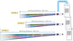
In this case, Optotune needed to engineer a solution to seamlessly adjust the working distance—the distance between the patient and the camera module—from a range of 350 mm all the way up to 650 mm, with an intermediate working distance of 420 mm. This level of adjustability and precision is a key advantage that liquid lenses hold over traditional fixed lenses. Unlike rigid, solid lenses that would require dramatic changes in their focal power (often in the range of +1 diopter to -1.7 diopters) to compensate for these working distance fluctuations, the liquid lens can achieve the necessary focus adjustments through only minimal changes to the curvature of the lens surface (see Fig. 3).
This adaptability is enabled by the inherent flexibility and dynamic nature of the liquid material comprising the lens, which allows it to fluidly morph its shape to rapidly fine-tune the focal characteristics as needed—without the mechanical complexity and limitations of moving parts that plague many traditional lens designs.
Using simulation, Optotune was able to meticulously model, test, and validate the optimal liquid lens parameters required to deliver this level of versatile, high-performance optical functionality.
The high-performance lens was engineered with exacting precision to meet and exceed the stringent optical quality standards set by Optotune, the manufacturer. The lens was required to have a distortion rate of less than 1.5%, but achieved an even lower distortion of less than 1% across all three tested distances. This is a remarkable feat of optical engineering, because minimizing distortion is notoriously challenging, particularly within the corners of the frame where it tends to be most pronounced.
In addition to the control of distortion, the lens also delivered outstanding relative illumination—a measure of how evenly light is distributed across the image sensor. The target specification called for a relative illumination greater than 70%, but the lens surpassed it and achieved a full 95% relative illumination—even within the far corners of the frame. This ensures a beautifully uniform brightness and color balance from edge to edge, without the vignetting or darkening that can plague lower-quality optics.
Figure 4 offers a visual representation of the lens performance, with the orange dots indicating the nominal quality targets Optotune aimed to reach. The red dots, in turn, show the minimum acceptable production quality. Remarkably, the actual performance of the lens, represented by the nominal design, is situated well above even the target specifications—which demonstrates the engineering team's commitment to creating an exceptionally high-caliber, distortion-free lens with outstanding illumination characteristics.
Advantages of liquid lens technology
The integration of liquid lenses into melanoma detection devices offers several advantages:
Dynamic focusing. Liquid lenses allow rapid adjustments to focus on different layers of skin lesions, which enables detailed examination of surface features and deeper structures.
Compact design. Liquid lenses are lightweight and compact, which makes them ideal for handheld devices used for clinical settings.
Real-time imaging. With liquid lenses, clinicians can capture high-quality images in real-time to facilitate immediate assessment and decision-making.
Cost-effectiveness. Liquid lens technology offers a cost-effective solution compared to traditional optical components, which makes it accessible to a wider range of healthcare providers.
Future directions
Advancing technologies, including AI-powered image analysis and miniaturized wireless devices, hold immense potential to revolutionize the detection and management of melanoma in the future.
Liquid lens technology is spearheading the critical effort of early melanoma detection. By enhancing clinical imaging capabilities, these innovative lenses empower clinicians to identify melanoma in its earliest, most treatable stages to improve patient outcomes. With continued advancements through collaborative innovation, the future of melanoma detection and treatment looks increasingly promising.
About the Author
Tomas Zvolensky
Tomas Zvolensky is marketing manager at Optotune Slovakia (Trnava, Slovakia).


