Schmidt telescope, scallop eyes transform microscope objectives
A team at the University of Zurich (UZH; Switzerland) developed multi-immersion Schmidt objectives to produce images with mirrors instead of traditional lenses. The influence? Scallop eyes where light passes through a lens-like structure and two retinas layered on top of each other before hitting a curved mirror in the back of the eye, which reflects it back onto the retinas (see video).
The overall basis lies in the Schmidt telescope developed by optician Bernhard Schmidt in the 1930s, which involves both the reflection and refraction of light. In a Schmidt telescope, a spherical primary mirror receives light that has passed through a correcting plate (a thin aspherical lens). It compensates for image distortions such as spherical aberrations that are produced by the mirror.
Conventional obstacles
Conventional microscope objectives are complex in design, requiring numerous lenses made from different types of glass, all perfectly aligned, to achieve the highest image quality. This approach is both costly and inefficient.
Biological imaging is increasingly relying on clearing techniques for larger (thicker) tissue, organ, and entire organism samples to make them optically transparent. The researchers say most clearing techniques use immersion media that’s incompatible with conventional microscope objectives. Imaging such samples requires objectives that combine a large field of view, according to the team’s study—published in Nature Biotechnology—as well as a long working distance and high numerical aperture, so consistency with a range of immersion media is ideal.
Even compatible, lens-based objective setups can be used with only one immersion medium, such as water or air. This means different objectives are needed for different types of samples—another pricey and inefficient detail.
The UZH team’s Schmidt objective overcomes these obstacles. It features a spherical mirror and an aspherical correction plate, providing the multi-immersion capability and making it suitable for all standard media. This is a key advantage of the objective.
“No lens-based objective has such capability,” says “amateur astronomer” Fritjof Helmchen, a professor of neuroscience at UZH and co-director of the school’s Brain Research Institute. “This feature is particularly important in view of the diverse media used in modern tissue clearing protocols.”
In their work investigating the use of mirrors instead of lenses for microscope objectives and operating with various immersion media, Helmchen’s team used a Schmidt telescope with a liquid immersion medium. This produced high-quality images, demonstrating its adaptability. Proving the versatility of their Schmidt objectives, the researchers imaged neural activity of zebrafish larvae in vivo using several media including air, water, benzyl alcohol/benzyl benzoate, dibenzyl ether, and ethyl cinnamate.
What’s next?
The researchers agree the Schmidt objective has far-reaching potential, namely in light-sheet microscopy, a fluorescence imaging technique that uses a sheet of laser light to illuminate a thin slice of a sample. It has become more widely used for biological imaging because the process is quick and nondestructive.
“A major next step will be to adapt the optical system so the Schmidt objective can be used in a light-sheet microscope setup,” Helmchen says. “This would avoid the costs of multiphoton microscope setups and provide even simpler and faster 3D acquisition.”
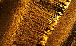
The Schmidt objective shows potential for several bioimaging applications, including neurology, pathology, and histology (see figure). It could be used for in vivo imaging applications, as well.
“This technology advances the high-resolution analysis of large tissue blocks,” says Helmchen, citing mainly anatomy, but also other applications requiring functional analysis. Further developments following the ideas of the Schmidt objective—for example, mirror-based designs tailored for 3D histology of large, cleared tissue blocks—have potential in the long term “to even transform tissue analysis on a larger scale.”
Physiological processes in most tissues are organized in a complex 3D manner, particularly in the nervous system, he adds, so “these advances promise to foster new insights into the functional organization and operating principles of diverse organs and tissues. As neuroscientists, we are excited to now have tools at hand with which we can better understand the distributed, large-scale organization of neural networks as the basis for information processing in the brain and adaptive animal behavior.”
About the Author
Justine Murphy
Multimedia Director, Digital Infrastructure
Justine Murphy is the multimedia director for Endeavor Business Media's Digital Infrastructure Group. She is a multiple award-winning writer and editor with more 20 years of experience in newspaper publishing as well as public relations, marketing, and communications. For nearly 10 years, she has covered all facets of the optics and photonics industry as an editor, writer, web news anchor, and podcast host for an internationally reaching magazine publishing company. Her work has earned accolades from the New England Press Association as well as the SIIA/Jesse H. Neal Awards. She received a B.A. from the Massachusetts College of Liberal Arts.

