Microscope component replacement improves photoacoustic imaging

Photoacoustic microscopy, an in vivo tissue imaging method based on the formation of sound waves that follow light absorption in a material sample, is an efficient way to see deep inside the body without contrast agents. But capturing super-resolution images with this technique remains challenging, as it’s difficult to focus light into a tiny, single point with just a lens.
Researchers at South Korea’s Pohang University of Science and Technology (POSTECH) are working on a way to overcome this challenge with a proof-of-concept system that replaces the lens with optical fiber. Their lens-free super-resolution system involves shear-force photoacoustic microscopy (SF-PAM) using a shear-force detection scheme and a tapered fiber (see Fig. 1 and video).

“Based on shear force detection, this system could analyze the optical absorption and scattering properties of samples as well as mechanical and morphological properties such as topography and viscosity,” says Byullee Park, a postdoctoral research associate in POSTECH’s Convergence IT Engineering department who co-led the study.
With the SF-PAM system, photoacoustic waves are generated in the near-field using an uncoated tapered fiber with an aperture size of 100 ± 25 nm. The tapered fiber is firmly attached to a quartz tuning fork (QTF), which is a piezoelectric dither, maintaining a stable distance of several tens of nanometers from the sample according to shear-force control schemes using a z-feedback loop of a phase-locked loop (PLL) system.
This ensures the same field energy and resolution by holding the probe at a constant distance along with the surface height of the sample during scanning.
For photoacoustic signal detection, the researchers positioned a customized high-frequency ultrasonic transducer diametrically opposite the fiber to the sample in the middle. To prove the feasibility of the system, Park says the light-field simulations are performed in the same structure as the tapered fiber; this field is applied to the photoacoustic simulation. A gold microlattice structure sample was then fabricated, and the photoacoustic imaging capability was demonstrated via 2D scanning.
In the study, the researchers’ SF-PAM system was able to acquire images with a resolution of 1 ± 0.3 μm, which is enough to clearly image cells, including red blood cells (see Fig. 2).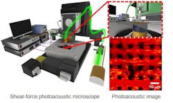
In a conventional photoacoustic microscope, light collects in the lens at a certain distance between the sample and the light source. But limitations of the lateral resolution and optical diffraction limit make it difficult to collect light at a single point in the lens. The researchers’ new approach side-steps the diffraction issue. Tapered, non-coated optical fiber that’s just tens of nanometers in diameter allows the distance between a sample and the light source to stay in the near-field range, where diffraction doesn’t occur.
The system aims to overcome other challenges as well, Park notes, including the need for a new direction in the field of super-resolution PAM and the need for a comprehensive nanoscale surface analysis system.
“This study is valuable as a proof-of-concept study aimed at super-resolution near-field scanning PAM in the future,” he says. “This is the cornerstone for obtaining super-resolution near-field scanning photoacoustic images through the development of non-metallic coating fibers.”
The system could potentially be used to study how diseases including cancer and cardiovascular disease form, which could lead to more effective treatments.
About the Author

Justine Murphy
Multimedia Director, Digital Infrastructure
Justine Murphy is the multimedia director for Endeavor Business Media's Digital Infrastructure Group. She is a multiple award-winning writer and editor with more 20 years of experience in newspaper publishing as well as public relations, marketing, and communications. For nearly 10 years, she has covered all facets of the optics and photonics industry as an editor, writer, web news anchor, and podcast host for an internationally reaching magazine publishing company. Her work has earned accolades from the New England Press Association as well as the SIIA/Jesse H. Neal Awards. She received a B.A. from the Massachusetts College of Liberal Arts.