Quantum technology could aid brain tumor surgery
From solar cells to atomic clocks for global positioning systems, quantum technology is making headway in a variety of applications. And now, brain tumor surgery could be added to the list.
Researchers from the German institutions Johannes Gutenberg University Mainz (JGU) and the Helmholtz Institute Mainz (HIM), working jointly on a German Federal Ministry of Education and Research-funded project, have developed a quantum sensor that could better ensure safe removal of brain tumors without harming healthy tissue that surround it or areas such as the motor cortex and nerve pathways (see video). The five-year project—DiaQNOS—kicked off in October, based on BrainQSens, a separate collaborative project in which JGU, medical doctors, and others developed highly sensitive magnetic sensors that enable improved medical diagnostics.
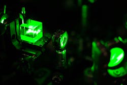
The JGU/HIM team’s new quantum sensor is a negatively charged nitrogen vacancy (NV) center—a point defect—in diamond (i.e., nanoscale magnetic field sensors confined in the diamond) that possesses an electronic-level structure, such as an atom, and can be optically initialized. Basically, notes DiaQNOS project coordinator Dr. Arne Wickenbrock, deputy section leader of the Budker Lab at JGU’s Institute of Physics and a researcher at HIM, they can be prepared and controlled in a certain spin-state and manipulated with microwaves.
“The high degree of control allows us to sensitively measure effects on the electronic-level structure,” he says. “NV centers are very sensitive to magnetic fields, and pressure, temperature, and rotation, as well.”
The DiaQNOS quantum sensor project has already allowed the team to improve the magnetic field sensor technology. Wickenbrock says that theoretically, it could register the brain’s magnetic fields (see figure). In a thin layer of diamond, many magnetic field sensors can exist, allowing the creation of a magnetic image of an object.
Wickenbrock explains that in the human body, nerve communication happens through electrical charges moving through nerve pathways. Each charge generates a magnetic field in the brain and throughout other parts of the body. The new quantum sensor detects and analyzes those fields.
Wickenbrock and fellow researchers in Mainz have been developing sensitive magnetometers based on NV centers in diamond since 2014. They’ve demonstrated that the sensitivity is sufficient enough to detect single-action potentials, but now, with DiaQNOS, the researchers aim to build an endoscopic magnetic imaging device, in addition to the quantum sensor.
“Not just a single sensor, but several to reconstruct an image of magnetic activity,” he says. “We can apply this to many things but together with partners at University Medical Center Freiburg, we would like to deploy it in the context of tumor surgery in the brain.”
The team’s goal is to build a device that allows them to measure the nerve activity in human brain tissue magnetically with a spatial resolution below 0.1 mm.
“Simply put, if the brain tissue is ok, nerves are firing periodically, which can be detected,” Wickenbrock says. “If the tissue is cancerous, this will change—either no activity or much different activity.”
The researchers may also be able to detect the tissue’s response to oscillating magnetic fields. This allows them to compute the conductivity, which itself can be used to distinguish tissue type and between cancerous and non-cancerous tumors. Once established as a tool, Wickenbrock says, the device could be used in many ways to image magnetic fields of objects, as well as the magnetophysiology of human organs.
“There is currently no magnetometer that combines the required sensitivity with this kind of spatial resolution, the 0.1 mm,” he says. “The hope is that the device will enable to diagnose cancer earlier and to extract cancerous material more precisely. This way, it can improve the outcome of neurosurgery, patient survival rate, and the amount of side effects for the patients.”
About the Author
Justine Murphy
Multimedia Director, Digital Infrastructure
Justine Murphy is the multimedia director for Endeavor Business Media's Digital Infrastructure Group. She is a multiple award-winning writer and editor with more 20 years of experience in newspaper publishing as well as public relations, marketing, and communications. For nearly 10 years, she has covered all facets of the optics and photonics industry as an editor, writer, web news anchor, and podcast host for an internationally reaching magazine publishing company. Her work has earned accolades from the New England Press Association as well as the SIIA/Jesse H. Neal Awards. She received a B.A. from the Massachusetts College of Liberal Arts.

