Imaging method detects damaged blood cells in real time
Mechanical devices such as artificial hearts and systems that assist blood flow are certainly medical marvels, but they are known to overstretch and even tear red blood cells. Now, researchers in Japan and Australia are working to prevent this currently unavoidable issue.
In a study published in Scientific Reports, the team, comprised of researchers from the Shibaura Institute of Technology (Japan) and Griffith University (Australia), notes that red blood cells (called erythrocytes) are regularly exposed to “shearing stress”—described as a force that causes layers or parts of a material/object to slide along each other in opposite directions—while circulating in the body. The stress is typically mild, as the red blood cells possess viscoelastic (flexible) properties, allowing them to accommodate shape changes in response to that stress. But this is not always the case.
Artificial hearts and other such mechanical devices often produce a stronger stress on the red blood cells, which can lead to rupturing and overstraining. Investigating such blood damage currently involves imaging the blood cells that are already damaged, according to the researchers, making it basically impossible to examine them in real time and (ideally) fix them. Doing so could help researchers to “better understand the process and aid in the development of markers to detect the onset of blood damage at a sublethal stage.”
The researchers note in their study that “given the need to minimize blood trauma within artificial organs, we observed [red blood cells] in supraphysiological shear through direct visualization to gain understanding of processes leading to blood damage.”
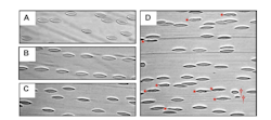
So, they’ve developed a new technique that could make it possible to advance that much-needed real-time study, and make the entire process more effective (see figure).
“Conventional image analysis methods are ill-equipped to detect subtle shape changes associated with morphological alterations observed in supraphysiological flow,” says researcher Nobuo Watanabe, an associate professor at Shibaura. “With our technique, it becomes possible to assess and detect these shape changes and damage.”
Essentially, the team’s method could allow for the early detection of the onset of such damage and potentially reverse it.
In their study, the researchers created a shear chamber and mounted it onto a microscope. To achieve this feat, the team used a custom-built shear chamber mounted on a high-speed camera-mounted microscope to image the onset of red blood cells. Blood samples from two healthy adult males were exposed to shear loads ranging from 10 to 60 pascals (Pa). This allowed them to detect abnormalities and subsequently adjust the imaging system they created “to improve asymmetry detection.” The researchers discovered that prolonged exposure to the larger stress load made the red blood cells “unstable, [they] changed shape, and even disintegrated into smaller fragments.”
“With our technique,” Watanabe says, “it becomes possible to assess and detect these shape changes and damage. Our proposed strategy focuses on early onsetting functional markers, when blood damage may still be reversible. Integration of such functional assessment markers into the deployment of cardiovascular medical devices could help guide future clinical practice of artificial organs to enhance blood compatibility and improve patient outcomes.”
About the Author
Justine Murphy
Multimedia Director, Digital Infrastructure
Justine Murphy is the multimedia director for Endeavor Business Media's Digital Infrastructure Group. She is a multiple award-winning writer and editor with more 20 years of experience in newspaper publishing as well as public relations, marketing, and communications. For nearly 10 years, she has covered all facets of the optics and photonics industry as an editor, writer, web news anchor, and podcast host for an internationally reaching magazine publishing company. Her work has earned accolades from the New England Press Association as well as the SIIA/Jesse H. Neal Awards. She received a B.A. from the Massachusetts College of Liberal Arts.

