Superresolution brain imaging gets boost from novel tuning approach
The approximately 250 nm resolution theoretically achievable with an optical microscope is not nearly high enough to deeply examine fine cellular structures such as live neurons. While stimulated-emission-depletion (STED) microscopy allows deeper imaging into those live neurons at higher resolution, scientists have discovered a way to go even further.
Researchers at Université de Bordeaux (Bordeaux, France) have developed a novel calibration method they say is simple and effective, and can also provide more precise STED imaging and at higher tissue depths. Ultimately, the team was aiming to “validate a method based on adaptive optics to achieve an a priori correction of spherical aberrations as a function of imaging depth.”
The study, published in Neurophotonics, demonstrates the approach that analyzes and corrects “one of the main sources of systematic error in STED microscopy for biological samples,” which is the spherical aberration of the depletion beam.1
According to the team, led by Professor Dr. U. Valentin Nägerl, “when imaging a tissue sample at depths higher than 40 μm, the depletion beam suffers various types of defocusing and degradation (aberration) and loses its carefully crafted shape” that is vital with the STED method. The researchers specifically looked at spherical aberration, which they cite as “the biggest offender.”
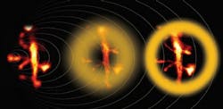
In the study, the team first measured the aberrations in a phantom sample of gold and fluorescent nanoparticles that was suspended in an agarose gel with a refractive index closely matching living brain tissue. Using a spatial light modulator, they applied corrective phase shifts; this provided clear visualization and allowed the researchers to quantify how the shape of the depletion beam became distorted as it penetrated deeper.
The imaging was achieved, according to the study, with a custom-built upright STED microscope that was based on pulsed excitation at 900 nm and pulsed depletion at 592 nm (see figure).
“Using our calibration strategy, we could measure neuronal structures as small as 80 nm at a depth of 90 μm inside biological tissue,” Nägerl says, “and obtain a 60% signal increase after correction for spherical aberration.”
Ultimately, the team found that the corrected STED images “captured the fine details of deeper neural dendrites much better than standard STED images.”
The researchers have found their “novel calibration process” to be “robust, straightforward to implement, and relatively inexpensive,” potentially making it easier to combine with standard lab practices. This could produce better results with STED microscopes, “so long as the prepared phantom sample matches the optical properties of the biological specimen.”
"Our approach is not limited to brain samples,” Nägerl says. “It could be adapted to other tissues with known and relatively homogeneous refractive indices, as well as other types of preparations, even potentially in the intact, live mouse brain.”
REFERENCE
1. S. Bancelin et al., Neurophotonics (2021); doi:10.1117/1.nph.8.3.035001.
About the Author
Justine Murphy
Multimedia Director, Digital Infrastructure
Justine Murphy is the multimedia director for Endeavor Business Media's Digital Infrastructure Group. She is a multiple award-winning writer and editor with more 20 years of experience in newspaper publishing as well as public relations, marketing, and communications. For nearly 10 years, she has covered all facets of the optics and photonics industry as an editor, writer, web news anchor, and podcast host for an internationally reaching magazine publishing company. Her work has earned accolades from the New England Press Association as well as the SIIA/Jesse H. Neal Awards. She received a B.A. from the Massachusetts College of Liberal Arts.

