Optoacoustic Imaging: Breast-cancer diagnostic tool can eliminate some physical biopsies
Early detection and diagnosis using ultrasound and/or x-ray mammography is a critical factor in successful treatment of breast cancer. However, these traditional imaging techniques only map morphology: the size, shape, location, and density of any growth inside the breast. An accurate determination of whether or not a mass is malignant still requires a physical biopsy, entailing patient discomfort, stress, recovery time, and cost. Typically, physicians err on the side of caution, meaning that as many as 71% of patients are subjected to biopsies of benign lesions,1 which can be particularly traumatic for women with masses located deep within the breast.
To help surmount this problem, engineers at Seno Medical (San Antonio, TX) have developed a new laser-based tool to improve the fidelity of noninvasive imaging, and in the process potentially spare many patients from these unpleasant procedures.
The tool, called the Imagio optoacoustic/ultrasound (OA/US) breast-imaging system, arose from the company’s desire to improve the quality of care for breast-cancer diagnosis by developing a noninvasive imaging method that would reveal growth morphology and physiology, while also providing a map of malignancy probability. Malignant growths exhibit anomalous blood circulation—they are highly vascularized to support their fast growth, and this growth also means that their blood content is typically more deoxygenated than surrounding normal tissue. To that end, the company created a laser-based system that is coregistered to a standard grayscale ultrasound, called Imagio, which can map both vascular density and local blood oxygenation.
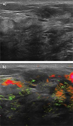
The system combines optoacoustic technology with B-mode ultrasound (see figure). A handheld, fiber-coupled unit similar to a conventional ultrasound probe is scanned over the breast. In addition to ultrasound interrogation, the unit illuminates the breast with nanosecond pulses of near-infrared (near-IR) light at two separate wavelengths. These are generated from a pair of custom-designed Q-switched lasers, one Nd:YAG and the other alexandrite. The lasers are coupled to the handpiece via a fiber bundle in the system umbilical.
How it works
“When tissue like a breast lesion absorbs a short laser pulse, it undergoes a very slight, but very rapid, temperature increase and physical expansion,” explains Sam Feldman, staff laser engineer at Seno Medical. “This energy is then released as a broad spectrum of acoustic waves, which can be recorded and spatially analyzed like the reflected ultrasound waves in a traditional ultrasound scan. The system alternates pulses at 1064 and 757 nm; the former is more preferentially absorbed by oxyhemoglobin, the latter by deoxyhemoglobin. In simple terms, the intensity of the acoustic signals indicates the degree of vascularization, and the ratio of the two signals indicates the extent of blood deoxygenation. In practical terms, there’s an enormous amount of proprietary knowledge involved in signal retrieval, image processing and reconstruction, and analysis.”
Generating high-energy nanosecond pulses dictates the use of a low-dispersion solid-state laser source based on bulk optics rather than fiber. The performance of optical resonators can be quite sensitive to optomechanical alignment. Moreover, this type of architecture inevitably includes numerous adjustable optical mounts, both intracavity and between the laser and the system output. These must all be immune to shifts because of the typical thermal drifts, vibrations, and shocks encountered as the Imagio system is moved and used in a clinic or hospital setting.
“Waiting for a breast cancer diagnosis is extremely stressful for most patients. Unexpected system downtime, which might cancel or delay procedures, runs counter to our mission and simply cannot be tolerated,” says Lisa Bichsel, marketing director at Seno Medical. For this reason, the company evaluated and compared the stability of several different types of optical mounts as a function of temperature and vibration. They ultimately selected monolithic flexures from Siskiyou (Grants Pass, OR) since these provided an order-of-magnitude better stability over the next-best mounts in their tests. Unlike conventional flexures based on plates and leaf springs, these mounts are machined from a single piece of metal to eliminate any movement between metal components because of a mismatch in thermal expansion or mechanical creep. Based on these tests, Siskiyou then worked with Seno to customize the exact form and fit needed for their Imagio system’s optomechanical arrangement.
Bichsel notes that the Imagio breast-tissue imager is CE-marked in Europe and is awaiting premarket approval (PMA) from the FDA for U.S. commercial availability.
REFERENCE
1. A. Vlahiotis et al., ClinicoEcon. Outcomes Res., 10, 157–167 (Mar. 26, 2018).
About the Author

John Wallace
Senior Technical Editor (1998-2022)
John Wallace was with Laser Focus World for nearly 25 years, retiring in late June 2022. He obtained a bachelor's degree in mechanical engineering and physics at Rutgers University and a master's in optical engineering at the University of Rochester. Before becoming an editor, John worked as an engineer at RCA, Exxon, Eastman Kodak, and GCA Corporation.