Light Sources for Cytometry: As cytometry expands, short wavelengths look sweet
"Ultraviolet (UV) lasers are important (if not common) fixtures on flow cytometers," writes William G. Telford, who heads up the Flow Cytometry Core Facility at the National Cancer Institute.1
And the importance of UV sources is increasing. This is because the trend in flow cytometry has been to study as many parameters as technology will allow. But spillover between fluorescent probes (that is, overlap in emission spectra of the light-activated dyes) complicates design with requirements for optical compensation, and thus threatens to limit the number of parameters researchers can investigate. So, UV presents an exciting window of opportunity.
Presentations at the 32nd Congress of the International Society for Advancement of Cytometry (CYTO 2017, Boston, MA; June 10-14) described the work that Telford and coauthors did to evaluate the application of 320 nm laser excitation—and separately, deep-UV (below 300 nm)—in cytometry.2
Moving beyond niche
The researchers explained that UV excitation has been a niche area in cytometry. But as flow cytometry has expanded, demand has grown for additional fluorescent parameters. The recent introduction by Becton Dickinson (BD; Franklin Lakes, NJ) of its BD Horizon Brilliant Ultraviolet (BUV) fluorochromes was a significant factor in opening up the UV range.
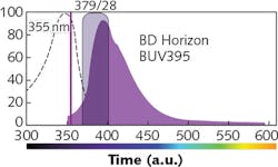
BD introduced the BUV395 (see figure) to provide an alternative to the UV-excitable fluorochromes available at the time that "are so dim that they are not practical for immunophenotyping applications," according to the company. Designed for excitation by 355 nm laser light and detection with a 379/28 filter, BUV395 was welcomed by cell scientists not only for its provision of an additional color, but importantly because it promised minimal (if any) spillover into other detectors.
Traditionally, water-cooled argon- and krypton-ion lasers have provided UV excitation in flow cytometry. More recently, 355 nm frequency-tripled diode-pumped solid-state sources (DPSS) became available-but they are physically large compared to other solid-state devices, and have correspondingly hefty price tags.
Seeking alternatives
Telford has been working to make UV even more accessible—that is, to help scientists take advantage of the new capabilities without breaking the budget. And while BD has specifically said that BUV395 is not recommended for use with 375 nm lasers, his work with colleagues reports that smaller and cheaper near-UV laser diodes (NUVLDs) delivering 375 nm light often can successfully stand in for 355 nm sources. But since the 375 nm sources don't pass muster with very short-wavelength probes, he worked with commercial partners to evaluate the appropriateness of new 320 nm (UV) and 280 nm (deep-UV) lasers for flow cytometry.
Telford's work with industry colleagues at LASOS (Jena, Germany) shows that the former (320 nm), which comes close to the 325 nm wavelength of helium-cadmium sources used in early benchtop cytometers, is nearly as effective as 355 nm sources for exciting most of the BUV dyes. And, they say, it performed just as well as the 355 nm source in population analysis of Hoechst and DyeCycle Violet side stem cells in mouse hematopoietic tissue. The 320 nm source also did an excellent job of exciting the indo-1 probe, which is incompatible with NUVLD 375 nm sources.
Overall, they summarize, the 320 nm laser module makes a suitable substitute for conventional 355 nm sources, and its availability in a more compact form factor than current 355 nm units makes it compatible with the trend toward smaller instrumentation.
Deep-UV
What about even shorter wavelengths? Historically, flow cytometry has not approached the deep-UV—that is, 300 nm and below. Besides the issue of probes, there are two reasons for this, the researchers say. First, 193 to 280 nm lasers have been available, but traditionally have pulsed at frequencies too low for flow cytometry and were so large as to be impractical for cytometer design. Second, commercial cytometers have not included optical elements that transmit such low-wavelength light.
Now, Telford says, "All three situations are poised to change."
Probes requiring deep-UV excitation are already being developed. System designers are experimenting with mirrors and optics that will make systems compatible with deep-UV. And compact, continuous-wave (CW) or quasi-CW deep-UV lasers are also emerging.
Working with industry colleagues at Oxxius S.A. (Lannion, France) and RPMC Lasers (O'Fallon, MO), Telford reported on tests using a CW solid-state 280 nm source mounted on a stream-in-air BD Influx cell sorter. The researchers replaced the instrument's optics with enhanced-UV aluminum mirrors and UV-compatible fused-silica lenses to enhance transmission efficiency and light focusing. Because no sub-300-nm organic fluorochromes yet exist, they used quantum-dot (Qdot) conjugated reagents, which are no longer widely used in flow cytometry.
They compared the performance of the 280 nm laser with that of a 355 nm source at the same power level, and found the modified cytometer to be completely compatible with 280 nm light. It provided efficient excitation of Qdot-labeled cells (and as expected, Qdot excitation was better at 280 nm than 355 nm). Excitation of the BUV dye was at <50% at 280 nm than at 355 nm—but the researchers consider that this is useful because it could allow the use of 280 and 355 nm simultaneously to excite different fluorochromes. The 280 nm laser light did excite considerable autofluorescence in the 380–450 nm emission region, but overall, say the researchers, this work suggests that deep UV may be useful as a distinct excitation source that could augment standard UV in flow cytometry.
REFERENCES
1. W. Telford et al., Cytometry A, 91, 4, 314-325 (2017).
2. W. Telford et al., "Deep ultraviolet lasers for flow cytometry," Proc. CYTO (2017).
About the Author

Barbara Gefvert
Editor-in-Chief, BioOptics World (2008-2020)
Barbara G. Gefvert has been a science and technology editor and writer since 1987, and served as editor in chief on multiple publications, including Sensors magazine for nearly a decade.