Intact Tissue Imaging/Light-sheet Microscopy: Clearing of intact bone promises insight into hard tissue
An expansion of CLARITY—a technique for optical clearing of soft tissue—renders intact bones transparent, and enables 3D observation of stem cells within marrow. The California Institute of Technology (Caltech; Pasadena, CA) scientists who developed Bone CLARITY have collaborated with researchers at biotechnology giant Amgen (Thousand Oaks, CA) to use it for testing of a new osteoporosis-treatment drug.
"The need for methods that provide 3D information to study the bone has long been recognized," the Caltech team reports in a paper co-authored by colleagues at Massachusetts General Hospital/Harvard Medical School (Boston, MA) and Amgen.1 Different types of bone are characterized by specialized processes, which scientists have traditionally investigated using methods such as cell sorting and histology that provide zero- or two-dimensional data. And while quantitative 3D imaging is now possible (to determine volume and number of cells, for instance), the capable approaches are labor-intensive and do not allow visualization of structure. Methods such as serial sectioning do reveal structure at the tissue level, but do not provide cellular-level information and cannot be easily combined with methods that do. And besides, they are destructive.
The Caltech team isn't the first to attempt bone clearing, but existing techniques have been unable to maintain native fluorescence in reporter mouse lines, and have achieved only limited imaging depth, the researchers say—and recent advances in imaging depth require bisectioning of bones.
Reading bones with light
The Bone CLARITY platform involves three parts. First, there is the clearing, which is challenging because bone involves two distinct types of tissue-hard mineral and soft marrow. The team's previous attempts enabled light penetration of only 200 to 300 μm into tissue. But Bone CLARITY allows imaging depth of up to ~1.5 mm: continuous convective flow during the clearing process allows for whole-bone transparency, the use of amino alcohol reduces autofluorescence, and a custom procedure minimizes refractive index (RI) variations. These factors also maintain the endogenous fluorescence of osteoprogenitor stem cells and a signal-to-noise ratio (SNR) that facilitates detection of individual cells. As a bonus, the process involves no harsh organic solvents.
The light-sheet imaging system, which includes a custom-built microscope, enables fast, high-resolution imaging without damaging the fluorescence signal. It illuminates specimens from two directions, and the ability to choose direction can dramatically reduce light scattering. Finally, dedicated computational methods count the fluorescently labeled cells.
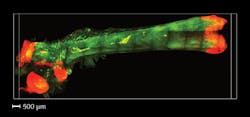
This three-part platform permitted visualization and quantification of the osteoprogenitors in a volume of mouse bone, and mapping of their 3D spatial distribution detection (see figure).
Testing treatment of osteoporosis
All healthy bone includes cells that build mass and those that break down aging tissue as part of a renewal cycle. The process is partially controlled by the stem cells in marrow called osteoprogenitors. "Because of the sparsity of the stem cell population in the bone, it is challenging to extrapolate their numbers and positions from just a few slices of bone," says postdoctoral scholar Alon Greenbaum, who is co-first author on a paper describing the work. And yet studying behavior of osteoprogenitors in their natural environment is considered critical for insight into diseases such as osteoporosis. According to Greenbaum, "there is a need to see inside intact tissue."
"Our collaborators at Amgen sent us a new therapeutic that increases bone mass," explains graduate student Ken Chan, the paper's other co-first author. The Amgen team was uncertain of the drug's effect on osteoprogenitors. "We reasoned that they might be increasing the proliferation of stem cells," Chan says.
Their study on mice demonstrated that "indeed there was an increase in stem cells with this drug," Chan says. "Monitoring stem cell responses to these kinds of drugs is crucial because early increases in proliferation are expected while new bone is being built, but long-term proliferation can lead to cancer."
Further application and development
Besides providing intraosseous insight, the technique promises to further understanding of how bones interact with the rest of the body. For instance, Bone CLARITY promises a new view into the cellular environment of the hemapoetic stem cells that are the focus of many labs studying aging. It could also be used to visualize environments that are difficult to access. For example, delicate lymphatic vessels that lie between the skull and brain are routinely damaged during mouse-brain removal, but Bone CLARITY could enable study while keeping the environment intact.
The team says that systemic delivery of antibodies and the use of small molecule staining methods could make the method more versatile. And to enable scaling to larger animals, development of stronger chelators, fast microscopes with very long working distances, and better ways to handle big data are required. Finally, the use of convolutional neural network (CNN) could further automate the precision and accuracy of the image processing pipeline.
REFERENCE
1. A. Greenbaum et al., Sci. Transl. Med., 9, eaah6518 (2017).
About the Author

Barbara Gefvert
Editor-in-Chief, BioOptics World (2008-2020)
Barbara G. Gefvert has been a science and technology editor and writer since 1987, and served as editor in chief on multiple publications, including Sensors magazine for nearly a decade.