Superresolution Microscopy: All-fiber illumination brings superior stability to STED
Stimulated emission depletion (STED) microscopy is one of the "super-resolved" fluorescence techniques honored with the 2014 Nobel Prize in Chemistry, recognized because of its demonstrated potential for advancing life sciences. Current implementations of STED—which uses Gaussian beams for excitation and donut-shaped modes carrying orbital angular momentum (OAM) as depletion beams—rely on free-space beam-shaping to precisely co-align these beams.
Researchers have long recognized that an optical-fiber-based version of STED would provide tremendous control of alignment tolerances, and that such an implementation could address engineering challenges including sensitivity of the point-spread function (PSF) to environmental perturbations, and high loss for mode conversion. Perhaps the greatest promise of a fiber-based setup, however, is endoscopic STED.
Earlier experiments with fiber-based STED have fallen short of such goals. But at CLEO 2016, Lu Yan of Boston University's Nanostructured Fibers and Nonlinear Optics Laboratory (Boston, MA) presented an all-fiber STED illumination system with low-loss and perturbation-tolerant vortex fiber modes excited by fiber gratings.1 Working with Siddharth Ramachandran and Poul Kristensen of OFS-Fitel (Brøndby, Denmark), Yan obtained naturally self-aligned PSFs for both excitation and depletion, with sizes and extinction ratios comparable to free-space implementations.
A compact, UV-inscribed, long-period grating (LPG) in the fiber with 99.7% conversion efficiency and <0.1 dB insertion loss excites the OAM modes, which the system stably propagates across the visible spectral range. The researchers tested the system's stability with a home-built STED microscopy system by obtaining PSFs for illumination with the Gaussian-shaped excitation beam, emitted as the fundamental LP01 beam of the fiber, as well as the ring-shaped STED beam, emitted as an OAM mode with L = ±1. They report having obtained mode purities of 19 dB and STED depletion beam extinction ratios of -20.5 dB while imposing a variety of fiber perturbations, including 10 loops of 6 mm radius.
The unprecedented stability results from a fiber design yielding enhanced effective index separation (Δneff >10-4) between the first higher-order vector modes (TE01, OAM [L = ±1], and TM01) across the visible spectral range. The researchers expect the fiber-based STED illumination system to compare favorably to state-of-the-art free-space STED microscopes.
Excitation and depletion light in same fiber
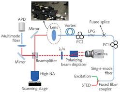
As described in the CLEO 2016 proceedings, the experimental setup (see figure) combines the excitation (λ= 632.8 nm; HeNe, Melles Griot) and depletion (λ= 776.34 nm; Ti:sapphire, Spectra-Physics) lasers into a single-mode fiber (SMF) with an inline fiber polarization controller (polcon) using a fused fiber coupler or a wavelength-division multiplexer. The researchers spliced the SMF to the vortex fiber, in which they inscribed a 31-mm-long tilted UV-LPG to generate OAM. They report that combined losses from the splice and grating are as low as 0.8 dB, and that splice optimization could enable additional improvements.
Following the grating, the team placed a second fiber polcon to control the content of spin-orbit-aligned OAM L = +1 and L = -1 modes, both of which generate a dark center under high-numerical-aperture (NA) focusing, and hence are suitable for STED. The output pigtail, which terminates about 5.1 m of coiled vortex fiber, is clamped using a bare fiber adapter (BFT1, Thorlabs). (The fiber is compatible with various termination options.) The beam output is then collimated using an objective (UPlanApo 10X, Olympus), and focused by a high-NA objective (UPlanSApo 60X/1.35, Olympus) onto a sample plane.
The team assessed mode purity using spatial interferometry, projecting the output of the fiber into two circular polarizations with a quarter-wave plate and polarizing beam displacer, and performing spatial Fourier analysis on the azimuthal intensity profiles. The researchers confirmed that, even with a 6 mm radius, the OAM mode remains greater than 98.7% (19 dB) pure. Sharper bends induce significant loss, but still perform better than the bend specifications for typical microscopes or endoscopes.
For bend radii of 6.5 cm and 6 mm, the team measured PSF for the excitation and depletion beams, respectively, by scanning (NanoMax-TS, Thorlabs) a sample of gold beads (150 nm Au nanoparticles, Cytodiagnostics) and detecting the scattered light. The BU researchers noted that the two PSFs are spatially well aligned in both directions: the full-width half-maximum (FWHM) of the excitation PSF is ~382 nm, and that of the dark center of the donut is ~235 nm.
"These values compare well with our theoretical estimates and are representative of similar measurements with state-of-the-art free-space systems," says Yan. He and his colleagues have measured extinction ratios (the ratios between intensity at the center and at the peak) as -17.6 dB and -20.5 dB, respectively—measurements that compare favorably to -13 dB, the oft-quoted desirable value for practical STED applications.
Going forward, the researchers plan to demonstrate full STED microscopy by collecting the fluorescence in the return beam path. They also anticipate integration of micro-optics such as gradient-index (GRIN) objectives to realize endoscopic STED imaging. And further, they say, production of a simplified, mechanically robust all-fiber STED system opens the door to realizing fiber versions of other microscopy techniques that exploit spatiospectral beam shaping.
REFERENCE
1. L. Yan, P. Kristensen, and S. Ramachandran, "All-fiber STED microscopy illumination system," CLEO 2016, OSA Technical Digest, paper SM4P.3 (2016).
About the Author

Barbara Gefvert
Editor-in-Chief, BioOptics World (2008-2020)
Barbara G. Gefvert has been a science and technology editor and writer since 1987, and served as editor in chief on multiple publications, including Sensors magazine for nearly a decade.