Stereoscopic vision generates 3D nanostructure images using X-rays
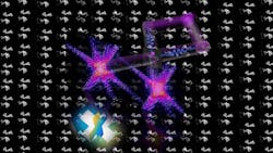
IMAGE: Two images of an object can be taken from two different directions using a single laser pulse, creating 3D X-ray images that enable rapid analysis of protein structures of viruses, for example. (Image credit: Hamed Merdji/CEA-Saclay)
Lensless microscopy with X-rays, or coherent diffractive imaging, is a promising approach. It allows researchers to analyse complex three-dimensional structures, which frequently exist in nature, from a dynamic perspective. Whilst two-dimensional images can already be generated quickly and in an efficient manner, creating 3D images still presents a challenge. Generally, three-dimensional images of an object are computed from hundreds of individual images. This takes a significant amount of time, as well as large amounts of data and high radiation values.
A team of researchers from Leibniz University Hannover and other universities has now succeeded in accelerating this process considerably. The researchers developed a method in which two images of an object can be taken from two different directions using a single laser pulse. The images are then combined to form a spatial image--similar to the human brain forming a stereo image from two slightly different images of both eyes. The method of computer-assisted stereoscopic vision is already used in the fields of machine vision and robotics. Now researchers have used the method in X-ray imaging for the first time.
"Our method enables 3D reconstructions on a nanometric scale using a single image which consists of two images from two different perspectives," says professor Milutin Kovacev from the Institute of Quantum Optics at Leibniz University Hannover, who is one of the co-authors of the study published in Nature.
According to the authors, the method will have a significant impact on 3D structural imaging of individual macromolecules and could be used in biology, medicine, as well as in industry. For example, the protein structure of a virus could be analyzed faster and with very little effort. The protein structure has an immense influence on the function and behavior of a virus and plays a decisive role in medical diagnoses.
The project was funded by Laserlab Europe, a consortium of European laboratories that aims to foster interdisciplinary laser research.
SOURCE: Leibniz University Hannover; https://www.uni-hannover.de/en/universitaet/aktuelles/online-aktuell/details/news/new-method-to-create-ultrafast-3d-images-of-nanostructures/
About the Author

Gail Overton
Senior Editor (2004-2020)
Gail has more than 30 years of engineering, marketing, product management, and editorial experience in the photonics and optical communications industry. Before joining the staff at Laser Focus World in 2004, she held many product management and product marketing roles in the fiber-optics industry, most notably at Hughes (El Segundo, CA), GTE Labs (Waltham, MA), Corning (Corning, NY), Photon Kinetics (Beaverton, OR), and Newport Corporation (Irvine, CA). During her marketing career, Gail published articles in WDM Solutions and Sensors magazine and traveled internationally to conduct product and sales training. Gail received her BS degree in physics, with an emphasis in optics, from San Diego State University in San Diego, CA in May 1986.