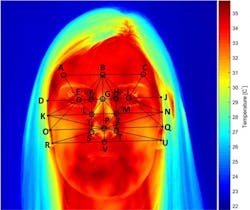
Researchers around the world are using infrared (IR) cameras to capture and record temperature variations on the skin for medical diagnostic purposes. By analyzing the images, researchers glean information on metabolic and vascular activity to recognize abnormal changes in physiology.
RELATED ARTICLE: Thermal imaging improves sports medicine and exercise research
Thermal imaging is used to study a broad number of diseases where skin temperature can reflect the presence of inflammation in underlying tissues or where blood flow is increased or decreased due to a clinical irregularity. Currently, many physicians use thermal imaging cameras to detect a number of medical conditions, such as arthritis, repetitive strain injury, muscular pain, and circulatory problems.
Relieving pain
Rheumatoid arthritis is a chronic autoimmune disease that can affect the hands, wrists, feet, knees, and shoulders. Arthritic joints tend to have higher temperatures than nonarthritic joints, so thermography can help physicians evaluate and monitor inflammation caused by the early stages of the disease. Read the full article at the SOURCE for more information.
Infectious for infrared
Infectious skin diseases are another area in which thermal imaging technology can be employed. At the Institute of Microbiology and Hygiene at Charité University of Medicine in Berlin, Angela Schuster and her colleagues have shown that IRT can assess a skin infestation called tungiasis, which causes inflammation of the skin. Tungiasis is caused by fleas that bite the skin surface before burrowing into the epidermis. The fleas then burrow deeper, into the upper dermis, to feed from blood vessels.
Thermographic images of the foot of a person with tungiasis were captured using a FLIR T660bx camera. The results showed that the temperature around a lesion caused by tungiasis was significantly higher than the median temperature of the foot. Researchers believe that the technique might be used to diagnose hidden and atypical manifestations of tungiasis and other infectious skin diseases prevalent in the tropics.
Second that emotion
Some researchers are trying to learn whether thermography can determine an individual’s emotional state. At the University of Granada, researchers have analyzed thermal differences between subjects who were exposed to photos of their loved ones and those shown images from the International Affective Picture System, a standardized database of pictures for studying emotion and attention.
The results showed that when an individual viewed an image of a loved one, specific regions around the body increased in temperature by up to 2 degrees C. Even more interesting, the thermographic system revealed that while passion increases temperatures around the hands and face, empathy (or the capacity to place oneself in another’s position) decreases temperatures, especially in the nose. Read the full article at the SOURCE for more information.
SOURCE: AIA Vision Online; https://www.visiononline.org/vision-resources-details.cfm/vision-resources/Thermal-Imaging-to-Diagnose-Disease/content_id/6938
About the Author

Gail Overton
Senior Editor (2004-2020)
Gail has more than 30 years of engineering, marketing, product management, and editorial experience in the photonics and optical communications industry. Before joining the staff at Laser Focus World in 2004, she held many product management and product marketing roles in the fiber-optics industry, most notably at Hughes (El Segundo, CA), GTE Labs (Waltham, MA), Corning (Corning, NY), Photon Kinetics (Beaverton, OR), and Newport Corporation (Irvine, CA). During her marketing career, Gail published articles in WDM Solutions and Sensors magazine and traveled internationally to conduct product and sales training. Gail received her BS degree in physics, with an emphasis in optics, from San Diego State University in San Diego, CA in May 1986.
