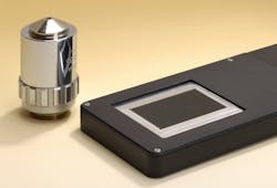
Jena, Germany--A microscope with a multichannel design that makes the scope compact and flat has been created by a group at the Fraunhofer Institute for Applied Optics and Precision Engineering IOF. Its purpose is to quickly and effectively detect melanoma on an area of a subject's skin in less than a second—so quickly that it can be held in the hand without blurring the resulting picture.
Array of micro-microscopes
The ultrathin microscope consists of many small imaging channels enable by microlens arrays. Each channel records a segment of the object at a 1:1 magnification. Each segment is roughly 300 x 300 µm2 in size and fits seamlessly alongside the neighboring segment; a computer program assembles the segments to generate an overall image. The difference between the technology and a scanning microscope is that all of the image segments are recorded simultaneously.
With an optical length of just 5.3 mm, the microscope is very thin. The microscope images to a resolution of five µm across a broad imaging area. "Essentially, we can examine a field as large as we want," remarks IOF group manager Frank Wippermann. "At five µm, the resolution is similar to that of a scanner."
The imaging system consists of three glass plates with microlenses both on top and beneath. The three plates are then stacked on top of one another. Each channel also contains two achromatic lenses, so the light passes through a total of eight lenses.
To fabricate the arrays, the scientists first coat the glass plate with photoresist and expose it to UV light through a mask. The plate is then etched, leaving cylinders of photoresist. Next, the researchers heat the glass plate; as a result, the cylinders melt, leaving spherical lenses. Working from this master tool, the researchers then generate an inverse tool that they use as a die. A die like this can then be used for mass production of the lenses.
The researchers have produced a first prototype and are showing it at Laser World of Photonics (May 23-26; Munich, Germany). With an image size of 36 x 24 mm2, the microscope can capture matchbox-sized objects in a single pass. It will be at least another one to two years before the device can go into series production, according to the researchers.
Subscribe now to Laser Focus World magazine; it’s free!
About the Author
John Wallace
Senior Technical Editor (1998-2022)
John Wallace was with Laser Focus World for nearly 25 years, retiring in late June 2022. He obtained a bachelor's degree in mechanical engineering and physics at Rutgers University and a master's in optical engineering at the University of Rochester. Before becoming an editor, John worked as an engineer at RCA, Exxon, Eastman Kodak, and GCA Corporation.

