Spectroscopy: Time-stretch spectroscopy STEAMs ahead
ROBERT R. THOMSON
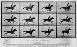
To clearly observe a time-evolving phenomenon, a measurement system with a temporal precision significantly higher than the phenomenon itself is required. A classic illustration of this concept is the 19th-century use of shuttered cameras by Eadweard Muybridge to photograph a galloping horse to resolve a debate over whether all four feet of the horse are ever fully off the ground during its gait (see Fig. 1). While this is indeed the case, it took a photograph to establish what the eye alone could not, since human vision does not have the required temporal resolution.
Technology has progressed significantly since the 19th century and we now have specialized high-speed scientific cameras that can deliver around a million frames per second (frames/s) with exposure times in the microsecond regime. But as incredible as these cameras are, they can only resolve processes that vary on the microsecond timescale.
Repetitive ultrafast events
So what if we want to observe even faster repetitive phenomena, such as those occurring on sub-nanosecond timescales? Fortunately, there are a variety of options available if the events are repetitive and always evolve in much the same way each time they occur.
Ultrafast gated intensified charge-coupled device (ICCD) cameras, for example, can provide gate times of hundreds of picoseconds and timing accuracies of a few picoseconds, enabling the user to build up a movie of the process by controlling the temporal position of the gate relative to the start of the process.
A wave of new, highly multiplexed detector array technologies is also emerging that incorporate hundreds of individual single-photon avalanche detectors (SPADs) into a single array, each of which has its own independent time-correlated single-photon counting (TCSPC) electronics.1 These arrays are now opening up exciting ultrafast imaging capabilities in areas that are intrinsically photon-starved, such as light-in-flight and through-body imaging.2, 3
In the area of ultrafast light-matter interactions, pump-probe techniques have been the workhorse tools for decades of research. In their most basic form, these techniques rely on using a leading pump pulse to initiate a process (such as the generation of excitons, or micro-explosions), together with a trailing probe pulse to probe the unfolding process.
To build up the time-averaged evolution of the process, this pump-probe measurement is repeated many times with different delays between the pump and probe. The power of this remarkably simple technique cannot be overstated. It was, for example, one of the key techniques that unlocked the field of ultrafast chemistry, finally resulting in the award of the 1999 Nobel Prize in Chemistry to Ahmed Hassan Zewail (the pioneer of the field), but it has also had broader impact in areas such as laser machining.4
Rare, non-repetitive ultrafast events
All the techniques outlined previously are suitable only for observing and measuring processes that are repetitive and do not vary significantly each time they occur. But when we wish to observe ultrafast phenomena that are rare and not repetitive, the photonic time-stretch (PTS) technique excels.
The most basic PTS method consists of three main steps. First, a short pulse of light is allowed to interact with the process of interest, such that the process imprints the desired information onto the spectrum of the pulse. Second, the spectrally modulated pulse is propagated through a dispersive medium (such as a length of dispersive optical fiber) so that the pulse spectrum is mapped onto its temporal profile-a process called wavelength-to-time mapping. Third, the temporal shape of the pulse is converted into a time-varying voltage signal using a photodetector, the profile of which is recorded using a real-time oscilloscope or similar instrument.
This elegant and powerful concept allows one to overcome the bandwidth limitations of current detector and digitizing technologies by stretching and slowing down the information contained within the spectrally modulated pulse at the end of the first step, to a timescale that can be sampled and recorded more effectively.
The key difference between the PTS technique and all the techniques and technologies described in the previous paragraphs is that PTS can be applied to phenomena that are not repetitive. In the case of PTS, the shutter speed of the measurement is determined by the temporal duration of the pulse at the beginning of the first step, and the frame rate is determined by the pulse repetition frequency, which can be hundreds of megahertz and extend even into the gigahertz regime.
Stretching time
The fact that PTS techniques can be used to measure and observe non-repetitive, rare phenomena makes it a truly enabling technology in many different areas where other techniques are too slow.
The basic techniques of optical wavelength-to-time-mapping and PTS were pioneered by the Jalali group at the University of California, Los Angeles (UCLA). In 2007, they used PTS principles to construct a real-time spectrometer capable of individually measuring the spectral properties of each pulse in a train of supercontinuum pulses. By doing so, they were able to demonstrate the existence of so-called optical rogue waves that are created during some nonlinear supercontinuum generation processes.5
In 2009, the same group reported the application of PTS methods to a breakthrough ultrahigh-speed imaging technique they named serial time-encoded amplified microscopy (STEAM).6 The concept behind STEAM is to use a spatial disperser to project different wavelength components within the pulse to different spatial positions across the system of interest. In this manner, it is clear that differences in the reflection amplitude across the system being probed will modify the spectrum of the reflected pulse (see Fig. 2).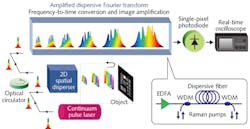
STEAMing ahead
The photonics community is currently on the cusp of new discoveries that explore the full range of PTS applications. Recent research results that merit mention include the first observations of exotic soliton explosions in a mode-locked fiber laser,7 and the first individual-pulse-resolved spectral evolution of a train of femtosecond pulses from initial noise fluctuations in a laser, providing insight into the processes that eventually lead to full mode locking.8
In more applied areas, one of the most exciting applications for PTS is STEAM imaging. In the biomedical sciences, for example, STEAM holds considerable promise for high-throughput cellular imaging, with researchers recently demonstrating the use of STEAM to develop a high-throughput imaging cell-based assay platform using a spinning digital video disc (DVD; see Fig. 4).9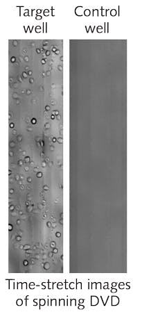
In this STEAM implementation, a simple diffraction grating is used to create a one-dimensional spectral shower on the DVD surface, rather than a two-dimensional spectral shower as used by Solli et al.6 To build up an image, the cells on the DVD are rapidly scanned past the spectral shower, at a velocity controlled by the spinning of the DVD. Impressively, the authors estimate that the overall imaging throughput of the system could be as high as 100,000 to one million cells per second, with potential applications in high-throughput screening in drug development and rare cancer cell screening.
Emerging technologies may also open up new opportunities for the PTS and its associated methods. One such example is the multicore fiber photonic lantern (pioneered by my colleague, Professor Tim A. Birks at the University of Bath in England)—a remarkable technology that enables the efficient interfacing of multimode signals with the single-mode cores of a multicore fiber.
In all the PTS implementations discussed here, the time-stretch itself is performed using a conventional single-core, single-mode fiber that restricts coupling efficiencies and throughputs when using multimode signals. As recently demonstrated by our group at Heriot-Watt University, in collaboration with the Birks and Henderson groups at the Universities of Bath and Edinburgh, respectively,10 the multicore fiber photonic lantern offers a route to highly multiplexed single-mode wavelength-to-time mapping using multimode states of light.
When used in combination with multi-pixel detector arrays, these devices open a route towards more efficient and versatile PTS systems exploiting the higher efficiencies of multimode optical systems. These capabilities could prove to be enabling in a variety of emerging areas, including new Raman spectroscopy modalities based on single-photon counting and wavelength-to-time mapping.11 These advances, and numerous others not discussed here, demonstrate the unique power and potential of PTS for a plethora of high-speed imaging and metrology applications.12
REFERENCES
1. J. Richardson et al., Proc. IEEE CICC 2009, 77–80 (2009).
2. G. Gariepy et al., Nature Commun., 6, 6021 (2015); doi:10.1038/ncomms7021.
3. M. G. Tanner et al., Biomed. Opt. Express, 8, 9, 4077–4095 (2017).
4. K. Sokolowski-Tinten et al., Phys. Rev. Lett., 81, 1, 224–227 (1998).
5. D. R. Solli et al., Nature, 450, 7172, 1054–1057 (2007).
6. K. Goda et al., Nature, 458, 7242, 1145–1149 (2009).
7. A. F. J. Runge et al., Optica, 2, 1, 36–39 (2015).
8. G. Herink et al., Nature Photon., 10, 321–326 (2016).
9. A. H. L. Tang et al., Biomed. Opt. Express, 8, 2, 640–652 (2017).
10. H. K. Chandrasekharan et al., Nature Commun., 8, 14080 (2017); doi:10.1038/ncomms14080.
11. Z. Meng et al., Proc. Nat. Acad. Sci., 112, 12315–12320 (2015).
12. A. Mahjoubfar et al., Nature Photon., 11, 341–351 (2017).
Robert R. Thomson was previously a Science & Technology Facilities Council (STFC; Swindon, England) Advanced Fellow and is currently professor of photonics at Heriot-Watt University, Edinburgh, Scotland; e-mail: [email protected]; http://phi.eps.hw.ac.uk.