From Ozcan's lab at UCLA: a smartphone fluorescence microscope and a lens-free cancer-detecting microscope
Aydogan Ozcan's lab at the University of California, Los Angeles (UCLA) has always been on the leading edge of microscope technology (see related links directly below). Now, Ozcan and his colleagues have created two of their newest examples: a lightweight, compact device that converts an ordinary smartphone into an advanced fluorescence microscope that detects and measures individual DNA molecules; and a small lens-free device that uses holography to detect cancer cells, doing the work of large pathology lab microscopes.1,2
RELATED: UCLA to begin testing lensless cell-phone-based microscope in Africa
RELATED: Holographic microscope is small, cheap, ready for use in remote areas
RELATED: COMPUTATIONAL IMAGING: Lens-free on-chip microscope is field-portable
RELATED: UCLA researchers create small Android/iPhone fluorescence device for common kidney tests
RELATED: LED-based lab-on-a-chip device screens for 170,000 molecules in blood
RELATED: Lensless optical microscope on a chip uses tomography to produce high-resolution 3-D images
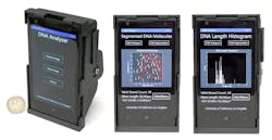
Smartphone fluorescence microscope
The mobile microscopy unit is an inexpensive, 3D-printed optical device that uses the phone's camera to visualize and measure the length of single-molecule DNA strands. The device includes an attachment that creates a high-contrast, dark-field imaging set-up using an inexpensive external lens, thin-film interference filters, a miniature dovetail stage, and a laser diode that obliquely excites the fluorescently labeled DNA molecules.
The sizing accuracy is better than 1 kilobase-pair (kbp) for 10 kbp and longer DNA samples imaged over a field of view of about 2 mm2.The UCLA researchers have reported the first demonstration of imaging and measuring the size of individual DNA molecules using a smartphone microscope. (Note: Ozcan's lab has done much work in the area of smartphone microscopes and other compact instruments useful for portable diagnostics and field use in developing countries.) The device also includes an app that connects the smartphone to a server at UCLA, which measures the lengths of the individual DNA molecules. The molecules are labeled and stretched on disposable chips that fit in the smartphone attachment. The application transmits the raw images to the server, which rapidly measures the length of each DNA strand. The results of DNA detection and length measurement can be seen on the mobile phone and on remote computers linked to the UCLA server.
"The ability to translate these and other existing microscopy and sensing techniques to field-portable, cost-effective and high-throughput instruments can make possible myriad new applications for point-of-care medicine and global health," says Ozcan.
Compact, lens-free pathology microscope
The lens-free microscope works by using a laser or LED to illuminate a tissue or blood sample that has been placed on a slide and inserted into the device. A cellphone-camera-style sensor array on a microchip captures and records the pattern of shadows created by the sample. The device processes these patterns as a series of holograms, forming 3D images of the specimen and giving medical personnel a virtual depth-of-field view. An algorithm color-codes the reconstructed images, making the contrasts in the samples more apparent than they would be in the holograms and making any abnormalities easier to detect.
The invention could lead to less expensive and more portable technology for performing common examinations of tissue, blood and other biomedical specimens. It may prove especially useful in remote areas and in cases where large numbers of samples need to be examined quickly.
Ozcan's team tested the device using Pap smears that indicated cervical cancer, tissue specimens containing cancerous breast cells, and blood samples containing sickle cell anemia. In a blind test, a board-certified pathologist analyzed sets of specimen images that had been created by the lens-free technology and by conventional microscopes. The pathologist's diagnoses using the lens-free microscopic images proved accurate 99% of the time.
Another benefit of the lens-free device is that it produces images that are several hundred times larger in area, or field of view, than those captured by conventional bright-field optical microscopes, which makes it possible to process specimens more quickly.
"While mobile health care has expanded rapidly with the growth of consumer electronics—cellphones in particular—pathology is still, by and large, constrained to advanced clinical laboratory settings," Ozcan says. "Accompanied by advances in its graphical-user interface, this platform could scale up for use in clinical, biomedical, scientific, educational, and citizen-science applications, among others."
Sources:
1. http://newsroom.ucla.edu/releases/ucla-engineers-first-to-detect-and-measure-individual-dna-molecules-using-smartphone-microscope
2. http://newsroom.ucla.edu/releases/lens-free-microscope-can-detect-cancer-at-the-cellular-level
REFERENCES:
1. Qingshan Wei et al., ACS Nano (2014); doi: 10.1021/nn505821y
2. Alon Greenbaum et al., Science Translational Medicine (2014); doi: 10.1126/scitranslmed.3009850
About the Author

John Wallace
Senior Technical Editor (1998-2022)
John Wallace was with Laser Focus World for nearly 25 years, retiring in late June 2022. He obtained a bachelor's degree in mechanical engineering and physics at Rutgers University and a master's in optical engineering at the University of Rochester. Before becoming an editor, John worked as an engineer at RCA, Exxon, Eastman Kodak, and GCA Corporation.