NIST developing skin-reflectance standards for hyperspectral imaging
After a successful non-human trial (see http://www.nist.gov/pml/div685/hyperspectral.cfm), researchers at the National Institute of Standards and Technology (NIST) have started gathering data on how human skin looks under various wavelengths of light in order to develop these badly needed standards.
RELATED ARTICLE: Optical ‘skin vibration’ signature yields biological parameters
Today, doctors who really want to see if a wound is healing have to do a biopsy or some other invasive technique that, besides injuring an already injured patient, can really only offer information about a small area. But hyperspectral imaging offers doctors a noninvasive, painless way to discriminate between healthy and diseased tissue and reveal how well damaged tissue is healing over a wide area. The catch? A lack of calibration standards is impeding its use.
According to NIST researcher David Allen, being sensitive to so many wavelengths means hyperspectral imagers can see many different things that humans can't see, including the amount of oxygen in human tissues, an indicator of healing. "The potential of the technology has been proven, but the problem is that researchers are simply lacking a way to assure consistent results between labs," says Allen. "Standards development has itself been hindered by a lack of human skin reflectance data, especially in the ultraviolet and short-wave infrared."
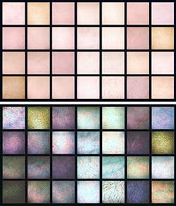
Catherine Cooksey, the project leader for the spectrophotometry program that establishes and maintains the national scale of reflectance, says that before we delve into what diseased tissue looks like hyperspectrally, we need to know what so-called "normal" tissue looks like. Furthermore, she says that they are looking to quantify the variability both within an individual and between individuals due to inherent biological differences. The initial NIST studies used 28 volunteer test subjects. The data collected included a photograph of the test area on the subject's forearm and three reflectance measurements of the test area.
"Skin reflectance varies due to skin pigmentation, tissue density, lipid content and blood volume changes," says Cooksey. "And few, if any, studies of skin reflectance have been done with an estimated measurement uncertainty that is traceable to NIST or any other national metrology institute. We need good data from a wide variety of sources, and for that we need the help of our colleagues in the community."
Once they collect enough data, the NIST researchers can feed it into NIST's Hyperspectral Image Projector (see http://www.nist.gov/pml/div685/grp04/hip.cfm), a device that creates hyperspectral scenes that have all the spectral signatures of the real thing--in this case, tissue in various stages of repair. Medical imaging technicians can then use these "digital tissue phantoms" to test their imagers' ability to discern among and detect different tissue types and conditions.
Those interested in helping to gather skin reflectance data should contact Allen ([email protected]) or Cooksey ([email protected]) for more information.
SOURCE: NIST; http://www.nist.gov/pml/div685/hyper-070114.cfm
About the Author

Gail Overton
Senior Editor (2004-2020)
Gail has more than 30 years of engineering, marketing, product management, and editorial experience in the photonics and optical communications industry. Before joining the staff at Laser Focus World in 2004, she held many product management and product marketing roles in the fiber-optics industry, most notably at Hughes (El Segundo, CA), GTE Labs (Waltham, MA), Corning (Corning, NY), Photon Kinetics (Beaverton, OR), and Newport Corporation (Irvine, CA). During her marketing career, Gail published articles in WDM Solutions and Sensors magazine and traveled internationally to conduct product and sales training. Gail received her BS degree in physics, with an emphasis in optics, from San Diego State University in San Diego, CA in May 1986.