A lot has changed since Galileo created the first microscope in 1609. Scientists began examining things like plant tissue, insects, and human blood cells, never realizing the advancements that would follow.
Today’s microscopy tools can take us past blood cells and tissues and into the atomic level. They’re offering new views and better understanding of diseases and viruses, prompting new approaches to treatment. Lasers have become standard tools in biomedicine—in many cases, they’ve replaced invasive surgeries for removing tumors and are increasingly used in laparoscopic procedures. Optical coherence tomography has greatly improved ophthalmology and optometry.
The biomedical field is evolving rapidly, and the advancement of these and other bio-optic and biophotonic tools is a prime example of that evolution.
Advances in photoacoustic imaging
There is an urgent need for brain imaging technology that can provide high-speed, high-resolution, large field of view, and functionality, all at the same time.
“These technical merits typically cannot be simultaneously met by a single imaging technology,” says Junjie Yao, an assistant professor of biomedical engineering at Duke University (Durham, NC).
He and his team in the school’s Photoacoustic Imaging Lab have developed a way to make this possible—ultrafast photoacoustic microscopy (UFF-PAM). The new technique can break all the limits of traditional brain imaging methodologies, according to Yao.
“The long-term goal is to apply this new technology to accelerate various brain research projects for fundamental neuroscience and provide important insights for treating neurological disorders such as stroke and Alzheimer’s disease,” he says.
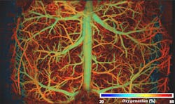
UFF-PAM allows researchers to engineer light and sound to probe the brain’s hemodynamic (blood flow) activities (see Fig. 1). Specifically, the technology listens to ultrasound waves generated by the biomolecules (e.g. hemoglobin in red blood cells) when excited by laser light. The imaging speed is accelerated by orders of magnitude by using state-of-the-art laser sources, scanning mechanisms, and ultrasound detection. A machine-learning technique is also incorporated into the signal processing to digitally enhance the image quality by intelligently filling the missing pixels of the image during the fast-scanning process.
Ultrafast photoacoustic imaging offers advantages over existing, traditional imaging methods, including its ability to meet all desired imaging parameters simultaneously. Existing methods may provide a subset of these parameters by sacrificing the others, “while our technology can do them all,” says Yao. “This provides a unique technical capability to examine the brain’s hemodynamics both at the large scale over the whole brain and the local scale at the single vessel level, which is not available by other imaging methods.”
The team’s research scaled several engineering hurdles. For the UFF-PAM to work properly and effectively, it requires an extremely high-speed laser to provide multiple optical wavelengths (colors) and more than 1 million shots per second. So, they developed a laser source based on the Raman-shifting effect—the energy difference between the incident (laser) light and the scattered (detected) light. Their new laser provides two optical wavelengths, at 532 nm and 558 nm, and is fast enough to support high imaging speeds.
They needed a scanning mechanism to steer the light beam from the laser while redirecting the resultant ultrasound waves to an ultrasound detector over a large field of view. To meet this requirement, they developed a polygon scanning system, which Yao says can provide the largest scanning range with the highest scanning speed to date in photoacoustic imaging.
He adds that another challenge for the team was avoiding “dumping too much light energy into the brain and inducing unwanted neuronal activities or tissue damage. We need to operate the imaging with sparse sampling, meaning we skip a large amount of image pixels to reduce the total light energy deposition. The missing pixels drastically reduce the final image quality.”
Creating a machine-learning technique to digitally make up the missing pixels and restore high image quality helped overcome this challenge.
“Almost all neurological diseases manifest changes to the brain’s vasculature and hemodynamic functions, which in turn have been highly informative for studying and treating the diseases in fundamental research and clinical practice,” Yao says. “Ultrafast photoacoustic microscopy is a useful tool to probe the disease mechanism, facilitate the diagnosis process, and provide monitoring and evaluating tool for developing new therapeutics.”
Cancer and disease detection
Biopsies are standard procedures used to detect and diagnose cancer; they can also unveil other conditions including autoimmune disorders. All types of biopsies are invasive in some way, from a percutaneous tissue biopsy, which involves inserting a small needle through the top layers of skin to collect cells, to surgical biopsies that often involve a deep incision to reach difficult-to-access cells. They’ve proven to be successful testing methods, but when it comes to diagnosing cancer or other conditions, patients and doctors should be armed with as many tools as possible. Now, a new device could soon be added to the arsenal.
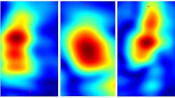
Researchers at the Stevens Institute of Technology (Hoboken, NJ) are developing a handheld device based on millimeter-wave imaging technology, which can see things through the skin even under poor visibility conditions (see Fig. 2). Negar Tavassolian, an associate professor at Stevens, says it’s a relatively mature technology already used in military and commercial applications, but not yet in biomedical ones.“The images we could get with this technology in biomedical applications didn't have high enough resolution,” she says. “The resolution was good enough for commercial and military, but not good enough for imaging tissue layers.”
Tavassolian and her team are exploring a different approach, similar to the one the TSA uses for airport security scanning. In millimeter-wave imaging for biomedical applications, the way millimeter-wave rays are reflected back from the skin depends on the type of tissue being investigated—cancerous or other suspicious tissue absorbs more rays than healthy tissue. Using antennas as well as antenna-radiating elements to monitor the reflection contrast and capture signals, the researchers produced a single ultra-high-bandwidth image. Ultimately, noise is reduced via this method, and high-resolution images of tissue can be captured within seconds.
But there are some challenges with this noninvasive technique, including getting adequate access to tissues. So, the new method would need to join with standard biopsies.
In some cases, “we will have to adjust our methods to what they do in the clinic with biopsies. We’re not trying to replace biopsies,” Tavassolian says. “For example, at one point we wanted to compare our images with histology images, but we couldn't access them.” So she and her team devised an algorithm to allow them to detect the presence of cancer.
Existing imaging devices for detecting cancer are large, expensive, and sometimes slow to produce images. The millimeter-wave imaging device developed by Tavassolian’s team is handheld and produces images quickly.
“The advantage of this kind of technology we’re working on is that we can use circuit technology, which is similar to what’s used in cell phones,” Tavassolian says. “So instead of what we have right now—a large tabletop device—we can reduce our device to the size of a cellphone. Then it's like a portable scanner.”
She cites dermatologists and surgeons as potential candidates for her team’s handheld imager, to be used in conjunction with standard biopsies. As millimeter-wave rays penetrate just a few millimeters into the skin, the new imaging device provides clear mapping of scanned lesions, something that’s not available via biopsy alone.
“In the mainstream dermatology market, when they're doing surgery, they want to remove part of the cancer,” she says. “But they don't know exactly where the markings of the tissue are with just a biopsy. They just trust their eyes. Our modules are more accurate.”
Grasping biological processes
Optical tweezers, a microscopy tool that uses a highly focused laser beam to hold and move microscopic and submicroscopic objects, can manipulate samples without mechanical interactions—an advantage over atomic force microscopy in bio-related studies; this can be done in 3D, and even inside samples (e.g. behind a layer of cells).
“The forces of optical tweezers and the extent of probe position fluctuation can easily be controlled with laser power,” says Alexander Rohrbach, a professor in the IMTEK Department of Microsystems Engineering at the University of Freiburg (Germany). “Optical tweezers can induce interaction processes, which do not occur spontaneously. This can be the unfolding and refolding of proteins on one side, but also the controlled binding of bacteria to immune cells on the other side.”
Rohrbach has been working with optical tweezers for more than two decades. Now, he and other IMTEK engineers are taking their study of this optical tool further to investigate how to more precisely control non-blind optical tweezers.
Traditionally, optical tweezers blindly grab at samples and objects, leaving those operating the microscope to manually position the tool. This can be challenging, particularly with objects too large to be rotated stably in fluid; the optical tweezers can’t get a solid hold. And force measurements and particle tracking remain difficult in inhomogeneous media, “where lots of scattering objects in the local environment disturb the optical trapping focus and the interference patterns for position detection,” Rohrbach says.
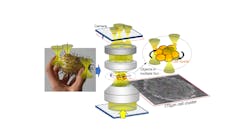
The researchers note in their study that while optical trapping forces are strong enough and related photodamage is acceptable, “the precise reorientation of large specimens with multiple optical traps is difficult, since they grab blindly at the object and often slip off.” The team’s approach allows them to localize and track regions with increased refractive index using several holographic optical traps with a single camera in an off-focus position (see Fig. 3).“Our study shows a way to find refractive index changes in large inhomogeneous media to find points where optical tweezers can apply efficient forces and torques to manipulate large objects some hundred microns in size,” Rohrbach says.
Optical tweezers combined with particle tracking open a new world of interaction measurements, allowing better understanding of the behavior of molecular binding partners as well as the binding and uptake behavior of cells with pathogens such as bacteria, viruses, or particulates.
“In this way, optical tweezers help a lot to understand the function and misfunction of cells and better understand molecular key players and processes in various diseases,” Rohrbach says.
While effective, optical tweezers technology hasn’t found its way into standard routine operations of research labs or in clinical applications just yet.
“Their operation is still more complicated than that of most microscopes and requires physicists or technicians,” Rohrbach points out. “The robustness of the systems and the reliability of the manipulation and measurement results need to be further improved in the future. Only when users can control their tweezing system in an adequate way will manipulations and measurements on a molecular scale become standard. Then users will discover with great excitement how fast interactions are on a molecular scale—motions and interactions much faster than imagined before.”


