Handheld MEMS-based OCT retinal scanner catches all info in one measurement

Dec. 23, 2013
Cambridge, MA--Researchers at the Massachusetts Institute of Technology (MIT), in collaboration with others at the University of Erlangen (Erlangen, Germany), Thorlabs (Newton, NJ), Praevium Research (Santa Barbara, CA), and Tufts University (Boston, MA), have created a handheld optical coherence tomography (OCT) imager that scans a patient’s entire retina in seconds.
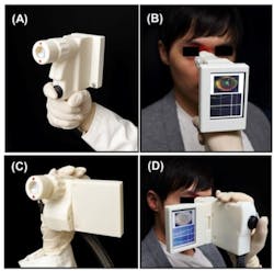
Power-grip-style (A-B) and camcorder-style designs (C-D) of a prototype OCT scanner are shown; both devices acquire 3D OCT images of the retina. (Credit: Biomedical Optics Express)
Cambridge, MA--Researchers at the Massachusetts Institute of Technology (MIT), in collaboration with others at the University of Erlangen (Erlangen, Germany), Thorlabs (Newton, NJ), Praevium Research (Santa Barbara, CA), and Tufts University (Boston, MA), have created a handheld optical coherence tomography (OCT) imager that scans a patient’s entire retina in seconds.1 The instrument, described in a paper published in the Optical Society's (OSA's) open-access journal Biomedical Optics Express, could aid primary-care physicians in the early detection of a host of retinal diseases including diabetic retinopathy, glaucoma and macular degeneration. Based on a 1060-nm-emitting vertical cavity surface-emitting laser (VCSEL) source from Praevium, the OCT instrument has a 350 kHz axial scan rate and produces a 10 µm axial resolution with a 3.08 mm axial imaging range; lateral range is either 6 x 6 mm or 10 x 10 mm. Scan time to capture a motion-corrected image is 1.4 s.
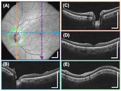
A high-definition OCT image of the retina allows clinicians to noninvasively visualize the 3D structure of key regions, such as the macula (region near the fovea) and optic-nerve head, to screen for signs of disease pathology. Shown here is a widefield view (A) as well as detailed vertical cross sections (B), (C), and (D), and a circular cross-section (E). Credit: (Biomedical Optics Express)

Although other research groups and companies have created handheld OCT devices using similar technology, the new design is the first to combine ultrahigh-speed 3-D imaging, a microelectromechanical systems (MEMS) mirror for scanning, and a technique to correct for unintentional movement by the patient. These innovations, the authors say, should allow clinicians to collect comprehensive data with just one measurement.

To deal with the motion instability of a handheld device, the instrument takes multiple 3D images at high speeds, scanning a particular volume of the eye many times but with different scanning directions. By using multiple 3D images of the same part of the retina, it is possible to correct for distortions due to motion of the operator’s hand or the subject’s own eye.

They tested two designs, one of which is similar to a handheld video camera with a flat-screen display. In their tests, the researchers found that their device can acquire images comparable in quality to conventional table-top OCT instruments used by ophthalmologists. The next step is to evaluate the technology in a clinical setting, says researcher James Fujimoto of MIT, an author of the paper. But the device is still relatively expensive, he adds, and before this technology finds its way into doctors' offices or in the field, manufacturers will have to find a way to support or lower its cost.

REFERENCE:


1. Chen D. Lu et al., Biomedical Optics Express (2014) http://dx.doi.org/10.1364/BOE.5.000293
Sponsored Recommendations

Request a quote: Micro 3D Printed Part or microArch micro-precision 3D printers

April 11, 2024
See the results for yourself! We'll print a benchmark part so that you can assess our quality. Just send us your file and we'll get to work.

Request a free Micro 3D Printed sample part

April 11, 2024
The best way to understand the part quality we can achieve is by seeing it first-hand. Request a free 3D printed high-precision sample part.

How to Tune Servo Systems: The Basics

April 10, 2024
Learn how to tune a servo system using frequency-based tools to meet system specifications by watching our webinar!

Precision Motion Control for Sample Manipulation in Ultra-High Resolution Tomography

April 10, 2024
Learn the critical items that designers and engineers must consider when attempting to achieve reliable ultra-high resolution tomography results here!

Voice your opinion!

To join the conversation, and become an exclusive member of Laser Focus World, create an account today!