Imaging sheds light on in-body movement of inhaled nanoparticles

Jan. 1, 2011
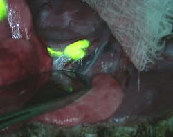
A collaborative effort from researchers at the Harvard School of Public Health, Beth Israel Deaconess Medical Center, Massachusetts Institute of Technology, and the German Research Center for Environmental Health using fluorescence imaging has resulted in a better understanding of the movement of potentially toxic nanoparticles within the body of a rat.

A collaborative effort from researchers at the Harvard School of Public Health (Boston, MA), Beth Israel Deaconess Medical Center (also in Boston), Massachusetts Institute of Technology (Cambridge, MA), and the German Research Center for Environmental Health (Neuherberg/Munich, Germany) using fluorescence imaging has resulted in a better understanding of the movement of potentially toxic—but potentially life-saving as well—nanoparticles within the body of a rat.

Using an intraoperative near-infrared fluorescence-imaging technique called fluorescence-assisted resection and exploration (FLARE), which simultaneously acquires 700 nm and 800 nm fluorescence images and color video images in real time, the researchers experimented using nanoparticles with a variety of diameters (up to 300 nm), shapes, and surface charges. The FLARE images revealed that inhaled nanoparticles smaller than 34 nm in diameter move from the lungs to the lymph nodes in as fast as 30 minutes, with even faster movement (a few minutes) for smaller particles. These smaller particles are also impacted by surface charge and can move quickly into the kidneys and bloodstream before being released in urine in as little as one hour from inhalation. The study has important implications for drug delivery and carcinogenesis, as nanoparticles could be optimized based on an understanding of where and how long they congregate in specific areas of the body. Contact Akira Tsuda at [email protected].
About the Author

Gail Overton | Senior Editor (2004-2020)

Gail has more than 30 years of engineering, marketing, product management, and editorial experience in the photonics and optical communications industry. Before joining the staff at Laser Focus World in 2004, she held many product management and product marketing roles in the fiber-optics industry, most notably at Hughes (El Segundo, CA), GTE Labs (Waltham, MA), Corning (Corning, NY), Photon Kinetics (Beaverton, OR), and Newport Corporation (Irvine, CA). During her marketing career, Gail published articles in WDM Solutions and Sensors magazine and traveled internationally to conduct product and sales training. Gail received her BS degree in physics, with an emphasis in optics, from San Diego State University in San Diego, CA in May 1986.

Sponsored Recommendations

Request a quote: Micro 3D Printed Part or microArch micro-precision 3D printers

April 11, 2024
See the results for yourself! We'll print a benchmark part so that you can assess our quality. Just send us your file and we'll get to work.

Request a Micro 3D Printed Benchmark Part: Send us your file.

April 11, 2024
See the results for yourself! We'll print a benchmark part so that you can assess our quality. Just send us your file and we'll get to work.

Request a free Micro 3D Printed sample part

April 11, 2024
The best way to understand the part quality we can achieve is by seeing it first-hand. Request a free 3D printed high-precision sample part.

How to Tune Servo Systems: The Basics

April 10, 2024
Learn how to tune a servo system using frequency-based tools to meet system specifications by watching our webinar!

Voice your opinion!

To join the conversation, and become an exclusive member of Laser Focus World, create an account today!